
Helicobacter pylori (H. pylori) and its significance in ENT Practice
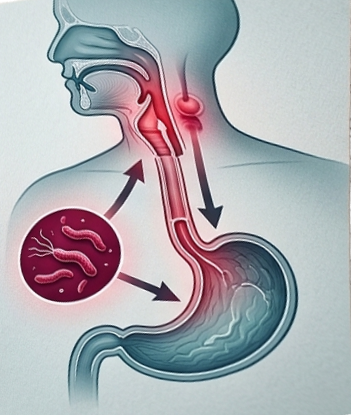
Overview
Helicobacter pylori was discovered in 1982 by Barry Marshall and Robin Warren (Nobel Prize 2005). It was previously thought that no bacteria could survive stomach acid.
It is now recognized as a major human pathogen, WHO Class I carcinogen, causing chronic gastritis, peptic ulcer disease, gastric adenocarcinoma and MALT lymphoma.
Significance in ENT: The upper aero-digestive tract is continuous with the stomach via reflux. H. pylori is now implicated in:
- Laryngopharyngeal reflux (LPR)
- Chronic laryngitis, supraglottitis, laryngeal cancer
- Chronic rhinosinusitis, nasal polyps
- Otitis media with effusion (via nasopharynx and Eustachian tube reflux)
- Halitosis, chronic tonsillitis, oral cavity lesions.
It survives in dental plaque, tonsillar crypts, adenoids, and nasal mucosa — acting as an extra-gastric reservoir
Definition and Structure
Definition:
Helicobacter pylori is a gram-negative, microaerophilic, spiral-shaped, motile bacterium that colonizes the gastric mucosa and extra-gastric sites of the upper aero-digestive tract.
Structure:
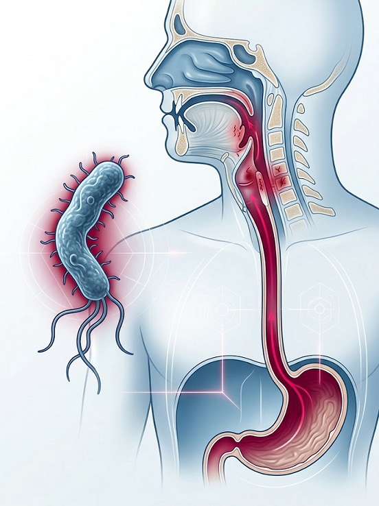
Figure : The shape of Helicobacter pylori
-
Shape:
S-shaped / curved rod, 2.5-4.0 µm x 0.5-1.0 µm, spiral
-
Motility:
4-6 sheathed unipolar flagella — allows corkscrew movement through mucus
-
Enzymes:
Abundant urease (converts urea to ammonia to neutralize acid), catalase, oxidase, mucinase, phospholipase
-
Virulence factors:
CagA (cytotoxin-associated gene A), VacA (vacuolating cytotoxin), BabA (adhesin), OipA
-
Microaerophilic:
Requires 5-10% O2, 5-10% CO2
-
Growth:
Fastidious, grows on chocolate agar, Skirrow's media at 37°C in 3-7 days
Epidemiology
-
Global prevalence:
∼50% of world population infected, ∼70-80% in developing countries (Ghana, Nigeria ∼70-90%), 20-40% in developed countries.
-
Who gets it:
Low socioeconomic status, overcrowding, poor sanitation, poor water supply.
-
How acquired:
- Feco-oral route (contaminated water/food)
- Oral-oral route (kissing, sharing utensils, mother to child)
- Gastro-oral route (vomitus reflux)
- Iatrogenic via endoscopes
-
Age:
Acquired in childhood, usually <10 years. Once acquired, persists lifelong if untreated. Highest seroprevalence in 40-60 years due to cumulative exposure.
-
Sex predilection:
Slight male predominance (M:F = 1.2:1), but no strong sex link. Males have higher rates of peptic ulcer and gastric cancer complications.
Pathology Caused by H. pylori
Gastric:
- Chronic active gastritis (antral predominant)
- Peptic ulcer disease (duodenal > gastric)
- Atrophic gastritis, intestinal metaplasia, dysplasia
- Gastric adenocarcinoma, MALT lymphoma
Extra-gastric / ENT-related:
-
Laryngeal:
Chronic laryngitis, laryngeal edema, contact granuloma, Reinke's edema
-
Pharyngeal:
Tonsillitis, Chronic pharyngitis
-
Nasal:
Chronic rhinosinusitis, nasal polyps
-
Otologic:
Otitis media with effusion, chronic suppurative otitis media
-
Oral:
Halitosis, aphthous ulcer, periodontitis, oral lichen planus
-
Esophageal:
GERD, Barrett's esophagus, adenocarcinoma (controversial)
Pathophysiology
-
Colonization:
Flagella + urease allow survival in acidic stomach and mucus of larynx/pharynx.
-
Adhesion:
BabA binds Lewis b antigen on epithelial cells.
-
Toxin release:
CagA injected via Type IV secretion system → disrupts cell junctions, activates NF-kB, IL-8 → chronic neutrophilic inflammation. VacA causes vacuolation and apoptosis.
-
Chronic inflammation:
Sustained IL-1, IL-6, TNF-alpha, oxidative stress → mucosal damage, atrophy → metaplasia → dysplasia → carcinoma sequence (Correa cascade).
-
Reflux mechanism in ENT:
Incompetent upper esophageal sphincter allows H. pylori -infected gastric juice to reach larynx/pharynx. The bacterium itself can colonize laryngeal mucosa (pH 6-7 favorable) and directly cause inflammation via same toxins.
-
Immune evasion:
Antigenic variation, inhibition of T-cell proliferation, survival inside macrophages.
H. pylori and Carcinoma in Upper Aerodigestive Tract
-
Gastric cancer:
WHO Class I carcinogen. Risk 2-6x increased. CagA+ strains highest risk.
-
Laryngeal cancer:
Emerging evidence. H. pylori DNA found in 35-60% of laryngeal squamous cell carcinoma specimens vs 10% controls. Mechanism: Chronic inflammation → DNA damage, CagA promotes proliferation, p53 mutation. Also synergistic with smoking/alcohol.
-
Hypopharyngeal and oropharyngeal cancer:
Similar chronic inflammatory pathway. Found in tonsillar tissue of cancer patients.
-
Esophageal adenocarcinoma:
Paradoxical — H. pylori may be protective against Barrett's via reduced acid, but CagA+ strains linked to cardia cancer.
Conclusion: Not yet proven as direct cause for laryngeal cancer like HPV, but strong co-factor.
H. pylori and Supraglottitis Leading to Upper Airway Obstruction
This is a rare but critical ENT emergency.
Mechanism:
- H. pylori colonizes supraglottis (epiglottis, aryepiglottic folds) via laryngopharyngeal reflux.
- Produces urease/ammonia + CagA → acute neutrophilic infiltration → edema of loose supraglottic mucosa.
- H. pylori also potentiates other organisms (H. influenzae, Streptococcus) — polymicrobial biofilm.
- Edema progresses rapidly → thumbprint sign on X-ray → stridor → complete airway obstruction.
Clinical picture:
Odynophagia, drooling, muffled voice, dyspnea, tripod posture. More common in adults with GERD/LPR and immunocompromised.
Management:
ABC — airway first. Do not attempt direct laryngoscopy in ward. ICU, IV steroids, PPI, H. pylori eradication + broad-spectrum antibiotics, ENT + anaesthesia standby for tracheostomy.
This is why persistent supraglottitis that fails to respond to standard antibiotics must be tested for H. pylori.
Symptoms - Specific and Non-Specific
Specific (Gastric/GERD/LPR related):
- Epigastric burning, dyspepsia, bloating.
- Hoarseness >3 weeks, chronic throat clearing
- Globus sensation, chronic cough (especially night)
- Halitosis (sulphur compounds from urease)
- Post-nasal drip, dysphagia
Non-Specific
- Nausea, loss of appetite, early satiety, weight loss
- Recurrent tonsillitis, nasal obstruction
- Ear fullness, hearing loss (otitis media with effusion)
- Fatigue, iron-deficiency anemia, idiopathic thrombocytopenic purpura
- Recurrent oral ulcers
Many patients are asymptomatic.
Signs of H. pylori Infection
- General: Pallor (anemia), low BMI
- Oral: Coated tongue, poor dentition, halitosis, tonsillar exudates
- Nasal: Purulent rhinorrhea, polyps.
- Ear: Retracted tympanic membrane, fluid level.
- Larynx (on laryngoscopy): Diffuse erythema, interarytenoid pachydermia, edema of vocal cords, contact granuloma, thick endolaryngeal mucus.
- Abdomen: Epigastric tenderness.
Investigations
Serology and Reliability
| Test | Sensitivity | Specificity | Reliability / Notes |
|---|---|---|---|
|
Serology IgG ELISA |
85%-90% |
75%-85% |
Detects exposure, NOT active infection. Remains positive 6-12 months after eradication. Poor for test-of-cure. Cheap, good for epidemiology screening. |
|
Stool Antigen Test (SAT) – monoclonal |
94% |
97% |
Detects ACTIVE infection. Good for diagnosis and test-of-cure. False negative if on PPI/bismuth. |
|
Urea Breath Test (13C-UBT) |
95 – 98% |
95 – 98% |
GOLD STANDARD non-invasive. Detects active infection. Most reliable. Expensive, needs equipment. |
|
Rapid Urease Test (CLO test) on biopsy |
90 – 95% |
95-98% |
Invasive, done during endoscopy. Quick. |
|
Histology (Giemsa/Warthin-Starry) |
95% |
≤ 98% |
Gold standard invasive. Also detects atrophy, cancer. |
|
Culture & PCR |
70 – 90% |
100% |
For antibiotic susceptibility. PCR can detect CagA and clarithromycin resistance. |
In ENT practice: Laryngeal brushings/biopsy for PCR, and UBT + stool antigen are preferred. Serology alone is unreliable for ENT decision making because it cannot differentiate past exposure from active laryngeal colonization.
Key rule: Stop PPI 2 weeks, antibiotics/bismuth 4 weeks before UBT or SAT.
Complications
- Peptic ulcer bleeding/perforation
- Gastric cancer, MALT lymphoma
- Iron deficiency anemia, Vitamin B12 deficiency, ITP
- Chronic LPR → laryngeal stenosis, vocal cord dysfunction
- Chronic rhinosinusitis resistant to treatment, otitis media → hearing loss, mastoiditis
- Recurrent supraglottitis → airway obstruction
- Failure of tonsillectomy/adenoidectomy due to reservoir reinfection.
Management
Triple vs Quadruple, 7 vs 14 days
Goal: Eradication >90%
-
Factors for choice:
Clarithromycin resistance >15% in region (Ghana/Nigeria high), prior macrolide exposure, penicillin allergy, cost, compliance.
First Line Therapy
(Maastricht VI / ACG 2024 Guidelines):-
Clarithromycin-based Triple Therapy (only if resistance <15% and no prior macrolide):
PPI (Omeprazole 20mg BD) + Clarithromycin 500mg BD + Amoxicillin 1g BD — 14 days
-
Bismuth Quadruple Therapy — PREFERRED first line in most developing countries:
- PPI BD + Bismuth subsalicylate 524mg QID + Tetracycline 500mg QID + Metronidazole 500mg TID — 14 days.
- Eradication rate: 90-95% vs 70-80% for triple therapy in high resistance areas -
Concomitant Quadruple (Non-bismuth):
PPI BD + Amoxicillin 1g BD + Clarithromycin 500mg BD + Metronidazole 500mg BD — 14 days
7 days vs 14 days:
7-day triple therapy: Eradication 70-75%, high failure, NO LONGER RECOMMENDED.
<br>
14-day therapy: Eradication 85-95%, significantly superior. All guidelines now recommend 14 days minimum. 10 days is intermediate but 14 is gold standard.
Second Line Therapy
If first line fails:If triple failed → Use Bismuth Quadruple for 14 days
If bismuth quadruple failed → Use:
- Levofloxacin-based triple: PPI + Amoxicillin 1g BD + Levofloxacin 500mg OD — 14 days
- OR Rifabutin-based: PPI + Amoxicillin 1g BD + Rifabutin 150mg BD — 14 days (reserve)
- Must do culture/susceptibility testing.
Levofloxacin resistance is rising — avoid as first line.
When to Repeat Serology Tests After Treatment
-
DO NOT use serology for test-of-cure
— IgG stays positive up to 1 year.
-
Use Urea Breath Test or Stool Antigen Test:
- Wait at least 4 weeks after finishing antibiotics
- Wait at least 2 weeks after stopping PPI
- If negative → eradication confirmed
- If positive → second line therapy needed
For ENT follow-up: Re - evaluate laryngoscopy after 6-8 weeks. .
Factors Affecting Response to Therapy
- Antibiotic resistance (clarithromycin, metronidazole, levofloxacin)
- Poor compliance (complex dosing, side effects)
- Inadequate PPI dosing (CYP2C19 rapid metabolizers)
- High bacterial load / biofilm in tonsils/dental plaque
- Smoking, alcohol
- Reinfection from spouse/family member (treat family)
- Not stopping PPI/antibiotics before testing
Conclusion, H. pylori is no longer just a gastroenterology organism. It is a significant extra-gastric pathogen in ENT practice, acting as a reservoir in tonsils, adenoids, dental plaque, and nasal mucosa, driving chronic laryngitis, LPR, rhinosinusitis, otitis media, halitosis, and potentially laryngeal carcinogenesis.
ENT surgeons must think beyond the stomach: chronic, recalcitrant ENT symptoms, especially LPR and supraglottitis, should prompt H. pylori screening using reliable tests (UBT/Stool Antigen, not serology alone). 14-day bismuth quadruple therapy is now the preferred first line in high-resistance areas, with test-of-cure at 4 weeks.
Eradication not only improves gastric disease but also reduces ENT recurrence and prevents progression to atrophy and malignancy.
Key Take - Home for ENT:
Treat the whole aero-digestive tract, not just the ear/nose/throat.
Share Post On:
Recent Posts
Categories


Get in Touch
Read doctor-produced health and medical information written for you to make informed decisions about your health concerns.
Address
Adenta
info@grovehealth.net
Working Hours
Mon - Friday @ 8am - 5 pm