Plummer-Vinson Syndrome / Paterson-Brown-Kelly Syndrome

Overview
PVS is a rare clinical triad: iron deficiency anemia + dysphagia + esophageal webs. Mostly seen in middle-aged women, though incidence dropped with better nutrition.
Historical Perspective + Names
British origin: Dr. Donald Paterson 1919 + Dr. Adam Brown-Kelly 1919, ENT surgeons in Manchester/UK, described dysphagia + postcricoid webs.
American origin: Dr. Henry Plummer 1912 + Dr. Porter Vinson 1919, Mayo Clinic physicians, linked it to anemia + oral/spoon nails.
Different names stuck by region. “Paterson-Brown-Kelly” is more common in UK, “Plummer-Vinson” in US.
Definition
Triad of :
1. Iron deficiency anemia,
2. Oropharyngeal / esophageal dysphagia,
3. Mucosal webs in upper esophagus / postcricoid area.
Epidemiology
Historically: Women 40-70y, Northern Europe / US early 1900s. Now rare in developed countries due to iron fortification. Still seen where iron deficiency is common. Prevalence ∼1-2% of IDA patients with dysphagia.
Pathophysiology
Which comes first?
Iron deficiency anemia comes first.
How anemia causes the web?
Chronic iron deficiency and reduced iron-dependent enzymes in esophageal mucosa and impaired epithelial regeneration + muscle function. Leads to mucosal atrophy, inflammation, fibrosis. Webs form as thin diaphragms of squamous mucosa + submucosa, usually postcricoid, 2-3 cm below cricopharyngeus. Also in oxidative stress + possible immune dysfunction.
Does syndrome include esophageal web or carcinoma?
-
1. Esophageal web:
Yes, it’s a core feature.
-
2. Esophageal carcinoma:
Not part of definition, but PVS is a premalignant condition. Risk of postcricoid/hypopharyngeal SCC is ∼3-15%. Need surveillance.
Symptoms & Signs
Symptoms
Dysphagia: progressive, painless, intermittent, for solids > liquids. Odynophagia if mucosal injury. Fatigue, glossitis, angular cheilitis, koilonychia/spoon nails, pallor, stomatitis.
Signs
Pallor, smooth/tender tongue, angular cheilitis, brittle/spoon nails, cervical lymphadenopathy if carcinoma develops.
Investigations
Laboratory:
CBC: microcytic hypochromic anemia. Low ferritin, low serum iron, high TIBC. B12/folate to rule out other causes.
Radiological:
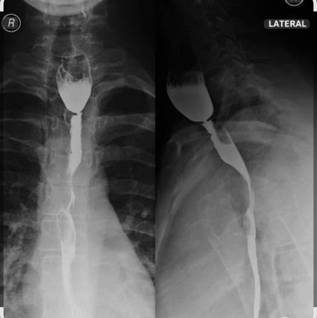
Barium esophagram / swallow study – “gold standard for esophageal web”. Shows thin, smooth, 2-3mm web in upper esophagus. Sensitivity ∼80-90%, false negatives if web torn by bolus.
Endoscopy:
Direct visualization + biopsy. High sensitivity/specificity for web. Allows dilation. False positives: mucosal folds. False negatives: small/partial webs missed if esophagus collapses.
Gold standard for esophageal web: Barium esophagram + UGI endoscopy together. Barium for anatomy, endoscopy for direct view + therapy.
Management
Medical:
Oral iron replacement 150-200mg elemental Fe/day + diet rich in iron/vit C. Treats anemia + often resolves dysphagia/web.
PPI if reflux present.
Surgical/Procedural:
Esophageal dilation – Savary-Gilliard dilators or balloon dilation under endoscopy. Done if iron alone fails. Success rate >90%.
Complications of condition
Oesophageal perforation from webs, aspiration pneumonia, weight loss / malnutrition, postcricoid SCC.
Complications of management
-
Iron:
GI upset, constipation, black stools, iron overload if misused.
-
Dilation:
Perforation 0.1-0.4%, bleeding, mediastinitis, recurrent web.
Conclusion
PVS = iron deficiency + dysphagia + esophageal web.
Reversible with iron, but carries malignant potential. Early diagnosis + iron repletion prevents webs + reduces cancer risk.
Share Post On:
Recent Posts
Categories
Get in Touch
Read doctor-produced health and medical information written for you to make informed decisions about your health concerns.
Address
Adenta
info@grovehealth.net
Working Hours
Mon - Friday @ 8am - 5 pm