
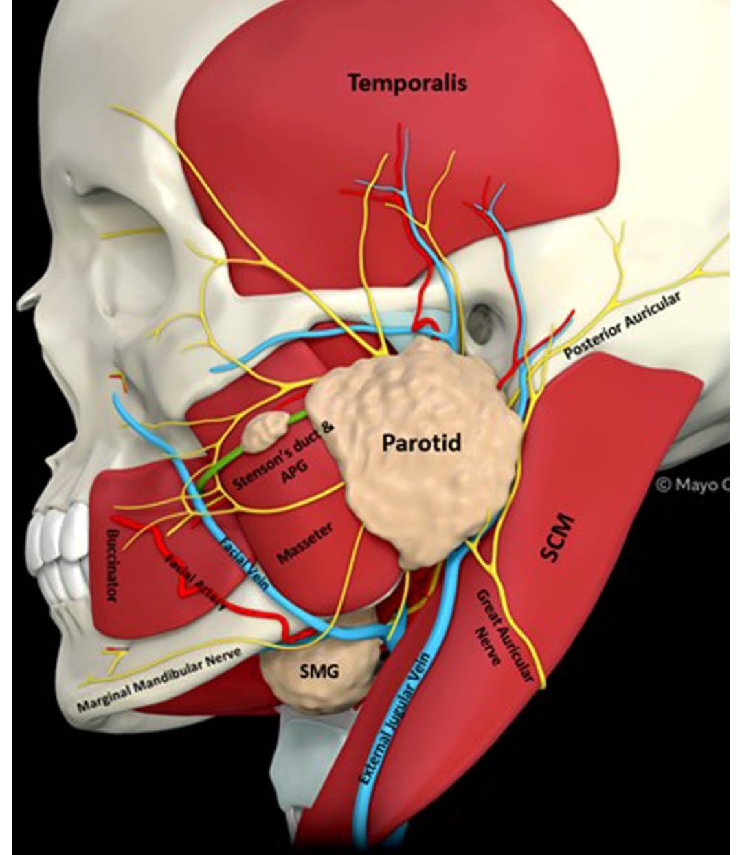
What is the Parotid Gland?
The parotid gland is the largest major salivary gland, paired, purely serous, irregularly wedge-shaped. Weight 25-30g. Contributes 25% of unstimulated but 70% of stimulated saliva. Most important surgically because facial nerve (VII) traverses it, dividing it into superficial and deep lobes – there is no true anatomical fascial septum, it’s a surgical division.
Enclosed in parotid fascia derived from investing layer of deep cervical fascia, forming a dense capsule.
Anatomy:
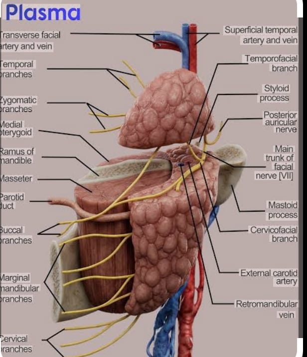
Figure 1: 3D Anatomy of the Parotid gland
Gross Anatomy & Location:
Located in parotid bed – retromandibular fossa, bounded: anteriorly – ramus of mandible, masseter; posteriorly – mastoid process, sternocleidomastoid; superiorly – external acoustic meatus, zygomatic arch; inferiorly – angle of mandible, posterior belly of digastric; medially – styloid process and its muscles, parapharyngeal space.
Surfaces: (lateral, anteromedial, posteromedial)
Processes: superficial lobe (80% of gland) and deep lobe/ retromandibular part (20%) connected by isthmus around facial nerve.
Duct - Stensen's duct:
5-7cm long, arises from anterior border, runs over masseter parallel to and 1cm below zygomatic arch, pierces buccinator at 90 degrees, opens opposite upper 2nd molar tooth at parotid papilla. Surface marking: middle 1/3 of line from tragus to midpoint between ala nasi and commissure of lips.
Relations:
Within gland: Facial nerve, retromandibular vein, external carotid artery (terminal branches), intraparotid lymph nodes
Superficial to gland: skin, superficial fascia, great auricular nerve (C2,C3), parotid fascia
Deep to gland: styloid process, styloid muscles, internal carotid artery, internal jugular vein, last 4 cranial nerves
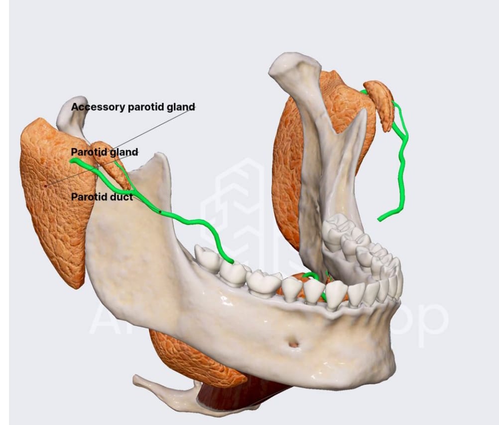
Figure 2: 3D view of the Parotid gland in relation to the mandible
Blood supply:
External carotid artery and its terminal branches – posterior auricular, superficial temporal.
Venous: retromandibular vein formed by maxillary + superficial temporal.
Lymph: Intraparotid and periparotid nodes -> upper deep cervical nodes.
Nerve supply:
Secretomotor parasympathetic: inferior salivatory nucleus -> IX -> tympanic branch -> tympanic plexus -> lesser petrosal nerve -> otic ganglion -> auriculotemporal nerve -> gland.
Sympathetic: superior cervical ganglion via plexus around external carotid – vasomotor.
Sensory: auriculotemporal nerve, great auricular nerve.
Motor through gland but not to gland: facial nerve
Layers from Skin to Gland (Parotidectomy approach):
- Skin
- Superficial fascia with platysma fibres, superficial musculoaponeurotic system (SMAS), great auricular nerve
- Parotid fascia (parotidomasseteric fascia) – dense investing fascia continuation
- Superficial lobe parenchyma
- Facial nerve plane (pes anserinus and branches)
- Deep lobe parenchyma
- Parapharyngeal space / stylomandibular membrane
Facial Nerve in Relation to Gland:
Facial nerve exits stylomastoid foramen, enters gland, divides into temporofacial and cervicofacial trunks, then into 5 terminal branches at anterior border: Temporal, Zygomatic, Buccal, Marginal Mandibular, Cervical – “Two Zebras Bit My Cheeks” – forming pes anserinus. Lies between superficial and deep lobes, more superficial inferiorly.
Landmarks to identify main trunk - Rules:
- Tragal pointer: 1cm deep and inferior to tip of tragal cartilage, nerve is 1cm below pointer.
- Tympanomastoid suture: 6-8mm deep to suture line.
- Posterior belly of digastric: Nerve lies 1cm superior to upper border of muscle, just deep to its attachment to mastoid groove.
- Styloid process: Nerve is lateral and superficial to styloid.
- Retrograde: Identify peripheral branch (marginal mandibular or buccal) and trace back.
Histology
Pure serous acinar gland. Serous acini with abundant zymogen granules, well-developed intercalated ducts and striated ducts. Fat cells increase with age. Contains intraparotid lymph nodes – important for metastasis and Warthin tumour origin from heterotopic ductal epithelium within lymph nodes.
Pathologies
Tumours:
80% of all salivary tumours occur in parotid, 80% benign.
Benign (80%):
- Pleomorphic adenoma – commonest overall (60-70% of parotid tumours) – 3-4:1 in superficial lobe
- Warthin tumour (papillary cystadenoma lymphomatosum) – 2nd commonest, almost exclusive to parotid, bilateral 10%, smoking association, male > female, from heterotopic tissue in lymph nodes
- Basal cell adenoma, Oncocytoma, Canalicular adenoma, Myoepithelioma
Malignant (20%):
- Mucoepidermoid carcinoma – commonest malignant in parotid, commonest in children too
- Adenoid cystic carcinoma – perineural invasion
- Acinic cell carcinoma
- Carcinoma ex pleomorphic adenoma
- Salivary duct carcinoma – aggressive, resembles breast duct carcinoma
- Adenocarcinoma (NOT OTHERWISE SPECIFIED- NOS), Squamous cell carcinoma, Lymphoma, Metastasis (skin SCC, melanoma to intraparotid nodes)
Sialolithiasis / Calculi
Less common – only 10-20% of salivary stones. Why rarer than submandibular:
- Stensen’s duct shorter, straighter, downward course with gravity
- Saliva serous, watery, less viscous, lower calcium, less mucin
- Faster flow, less stagnation
- More acidic pH, does not favor precipitation
Other Lesions:
- Cysts: first branchial cleft cyst (type I & II – closely related to facial nerve), parotid cysts in HIV
- Abscess: acute suppurative parotitis – ascending from oral cavity, elderly dehydrated post-op, Staph aureus
- Neurofibroma, hemangioma (commonest parotid tumour in children – infancy)
- Sialadenosis – non-inflammatory enlargement – diabetes, alcoholism, malnutrition
- Sialocele, fistula
Note on Sjogren's Syndrome:
Primary: autoimmune destruction of acini and ducts by CD4 T-cells. Secondary with connective tissue diseases (RA, SLE). Parotid is main gland affected for enlargement – bilateral, recurrent. Histology: benign lymphoepithelial lesion with epimyoepithelial islands. Diagnosis: Schirmer test, anti-Ro/La, lip biopsy of minor salivary glands (gold standard). Increased risk of non-Hodgkin MALT lymphoma 44-fold.
Investigations
-
Plain X-ray:
Rarely useful - only 40% parotid stones radio-opaque
-
Sialography:
Ductal architecture - sialectasis in Sjogren (punctate, cavitary)
-
Ultrasound:
First line for superficial lobe, differentiates solid vs cystic, guides FNAC. Can't see deep lobe well.
-
CT Scan:
Deep lobe, parapharyngeal extension, stones, abscess, bony involvement
-
Magnetic resonance imaging (MRI):
Gold standard for tumours - extent, deep lobe, facial nerve relation, perineural spread, distinction benign vs malignant (T1 hypo, T2 hyper, irregular borders). MR sialography for ductal system.
-
FNAC / Core biopsy:
Ultrasound-guided FNAC - Milan system for reporting
-
Sialendoscopy
Management of Pathologies
-
Sialadenitis:
Rehydration, antibiotics, sialogogues, gland massage
-
Sialolithiasis:
Conservative, sialendoscopic retrieval, extracorporeal lithotripsy, transoral duct approach, parotidectomy for intraglandular large stones with chronic damage
-
Benign tumours:
Superficial parotidectomy with facial nerve preservation is standard for superficial lobe pleomorphic adenoma. Enucleation is obsolete - high recurrence (40% due to pseudopods and capsule rupture). Extracapsular dissection for small superficial benign tumours in selected centres.
-
Malignant:
Total conservative parotidectomy (preserve nerve if not infiltrated) + neck dissection + post-op radiotherapy. Radical parotidectomy sacrificing nerve if directly invaded + nerve grafting.
-
Sjogren:
Symptomatic - artificial tears, saliva, pilocarpine, good oral hygiene, monitor for lymphoma
Parotidectomy
Step-By-Step Excision Of Parotid Gland
-
Indications:
Pleomorphic adenoma, Warthin, malignant tumours, chronic sialadenitis with fistula, intraglandular stones, first branchial cleft cyst, parotid lymphoepithelial cysts in HIV.
Types: Superficial parotidectomy, partial superficial, total conservative, radical.
Rule of 2s for Parotid:
- 2 main divisions of facial nerve
- 2 terminal arteries inside
- 2 veins - retromandibular and its branches
- 2 nerves to preserve - facial nerve and great auricular nerve (sensory) if possible
- 2cm below ear lobule - incision starts
Procedure - Superficial Parotidectomy (Blair's approach):
-
Anaesthesia:
General anaesthesia with endotracheal tube. No long-acting muscle relaxant after induction to allow facial nerve monitor. Eye protected, facial nerve monitor electrodes in orbicularis oculi and oris. Supine, head turned opposite, neck extended.
-
Skin incision:
Modified Blair incision - lazy S: starts preauricular in preauricular crease, 1mm anterior to tragus, extends to lobule, curves posteriorly around lobule 1cm below, then forward into upper neck crease 2cm below mandible towards hyoid. Alternative - face lift incision for cosmesis.
-
Elevation of flap:
- Incise skin, subcutaneous tissue.
- Elevate skin flap in plane of superficial fascia (between subcutaneous fat and parotid fascia/SMAS) anteriorly to anterior border of gland, superior to zygomatic arch, posterior to SCM, inferior to angle.
- Preserve great auricular nerve – if possible preserve posterior branch to earlobe to prevent numbness; anterior branch usually sacrificed.
-
Exposure of main trunk:
- Divide parotid fascia over SCM and mastoid.
- Retract SCM posteriorly, identify posterior belly of digastric muscle.
- Identify tragal pointer and tympanomastoid suture.
- Dissect antegrade: find main trunk 1cm deep to tragal pointer, just superior to digastric, 6-8mm deep to tympanomastoid suture.
- Use haemostat to spread along nerve direction.
-
Dissection of superficial lobe:
- Once trunk found, trace to pes anserinus.
- Dissect superficial lobe off facial nerve branches using fine mosquito and bipolar scissors – branch by branch from proximal to distal, lifting gland away from nerve.
- Retromandibular vein and external carotid branches ligated as encountered.
- Stensen’s duct ligated at anterior border if total superficial.
-
Deep lobe:
If needed for total conservative: after superficial lobe removed, identify branches again, dissect deep lobe from underneath nerve, careful of internal jugular and styloid.
-
Haemostasis:
Meticulous bipolar, Surgicel.
-
Drain:
Suction drain (10F) placed in parotid bed, exits posteriorly.
-
Closure:
SMAS/parotid fascia sutured to reduce Frey's, platysma 3-0 Vicryl, skin subcuticular. Pressure dressing for 48h.
Anaesthesia
Skin incision:
Anaesthesia
Exposure of main trunk
Dissection of superficial lobe
Deep lobe
Haemostasis
Drain
Closure
Figure 3: Steps of Excision Of Parotid Gland
Complications Of Parotid Surgery
-
Facial nerve injury:
Most feared. Temporary neuropraxia 15-30% (recovers weeks-months), permanent <5% in benign. Marginal mandibular most vulnerable.
-
Frey syndrome (gustatory sweating):
10-40% - parasympathetic aberrant regeneration to sweat glands, sweating/flushing while eating. Treat with antiperspirant, botulinum toxin.
-
Sialocele, salivary fistula:
Conservative - pressure, antisialogogues, scopolamine patch
-
Hematoma, seroma, infection
-
Great auricular nerve anesthesia:
Ear lobule numbness
-
First bite syndrome:
Pain in parotid on first bite after deep lobe surgery - loss of sympathetic innervation
-
Keloid / hypertrophic scar
-
Recurrence of
Pleomorphic adenoma if enucleation or spillage
-
Management:
Facial physiotherapy, eye care with tarsorrhaphy if needed, botox for Frey, evacuation of hematoma.
Methods of Managing Parotid Calculi
Conservative first. Sialendoscopy is gold standard – basket retrieval, laser fragmentation (Holmium:YAG). Extracorporeal shock wave lithotripsy. Transoral approach for distal Stensen’s. Parotidectomy only as last resort – risk to facial nerve.
Note on HIV infection of Parotid
Commonest salivary manifestation of HIV. Benign lymphoepithelial cysts – bilateral parotid enlargement (80%), multiple thin-walled cysts with lymphoid hyperplasia. Associated with DILS. CD8 infiltration, cervical lymphadenopathy. Diagnosis: HIV test, FNA – proteinaceous fluid with lymphocytes.
Treatment: HAART causes regression, repeated aspiration, sclerotherapy with doxycycline, low-dose radiotherapy, superficial parotidectomy for diagnosis/compression.
In conclusion,
Parotid gland is the largest salivary gland but surgically most challenging due to facial nerve traversing it. Unlike submandibular where calculi predominate, parotid predominance is neoplastic, mostly benign. Understanding facial nerve landmarks and preserving SMAS/fascia layers is key to safe parotidectomy with minimal morbidity.
Share Post On:
Recent Posts
Categories
RELATED POSTS
Get in Touch
Read doctor-produced health and medical information written for you to make informed decisions about your health concerns.
Address
Adenta
info@grovehealth.net
Working Hours
Mon - Friday @ 8am - 5 pm