
Overview Of The Submandibular Gland
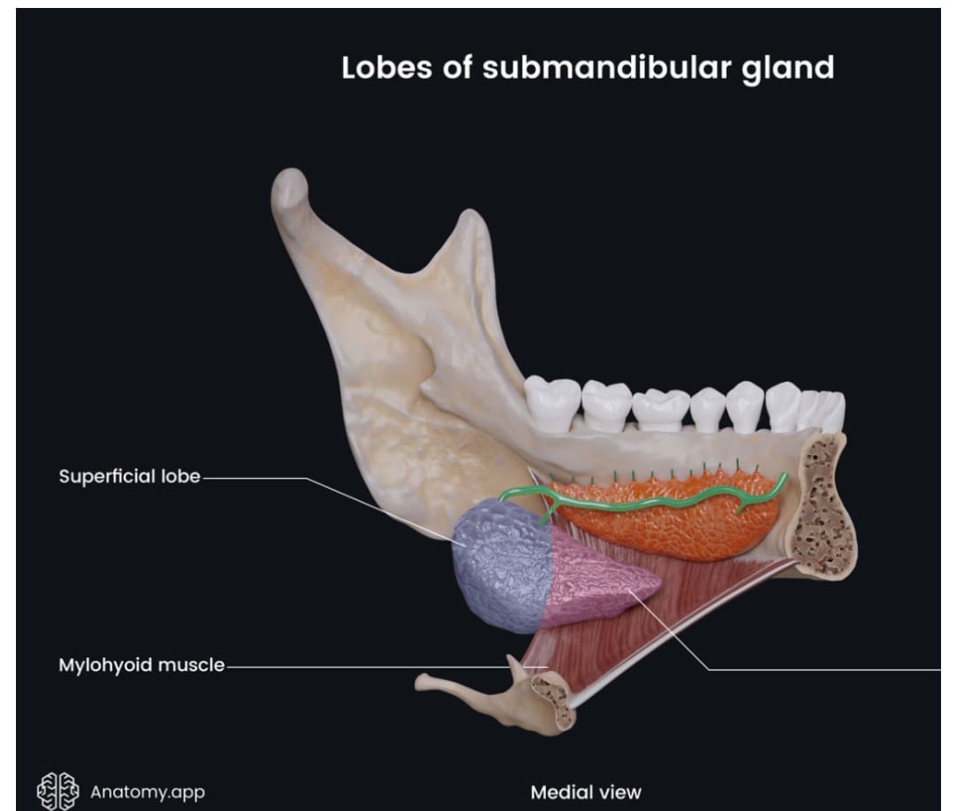
What is the Submandibular Gland?
The submandibular gland is the second largest major salivary gland, paired, mixed seromucous but predominantly serous. It contributes ∼60-70% of unstimulated whole saliva. Weight ∼7-16g.
It lies in the submandibular triangle, with a superficial and deep lobe continuous around the posterior border of mylohyoid.
Anatomy:
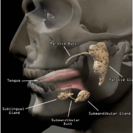
Figure 1: Anatomical relations of the major salivary glands
Gross Anatomy:
-
Superficial lobe:
Larger, fills most of submandibular triangle. Lies between mandible above and digastric muscles below.
-
Deep lobe:
Smaller, lies above mylohyoid, in floor of mouth, between mylohyoid and hyoglossus.
-
Duct - Wharton's duct (submandibular duct):
5cm long, arises from deep lobe, runs forward between mylohyoid and hyoglossus, then between sublingual gland and genioglossus, opens at sublingual caruncle beside lingual frenulum. Crosses lingual nerve.
Relations:
Superficial: skin, platysma, cervical branch of facial nerve
Medial: mylohyoid, hyoglossus, lingual nerve, hypoglossal nerve, facial artery
Posterior: tail of parotid, facial artery grooves its posterior surface
Lateral: mandible, medial pterygoid, facial vein
-
Blood supply:
Facial artery (submental and submandibular branches), lingual artery. Venous: facial vein, submental vein.
-
Lymph:
Submandibular lymph nodes -> deep cervical.
-
Nerve supply:
Secretomotor parasympathetic from superior salivatory nucleus -> chorda tympani -> lingual nerve -> submandibular ganglion -> gland. Sympathetic from superior cervical ganglion via facial artery plexus.
Layers from Skin to Gland:
- Skin
- Superficial fascia with platysma
- Deep cervical fascia (investing layer) forming capsule – splits to enclose gland.
- Superficial layer of deep fascia + marginal mandibular nerve + facial vein + cervical branch of facial nerve
- Glandular capsule (false capsule from investing fascia, true capsule from condensation)
- Gland parenchyma
Marginal Mandibular Nerve:
- Branch of facial nerve (VII)
- Runs in plane deep to platysma, superficial to investing fascia, often just below angle of mandible
- Relation:
Hayes Martin maneuver – nerve lies 1-2cm below mandible, but in 20% can be below. To preserve: incision 2 fingerbreadths (2-3cm) below mandible, elevate flap in subplatysmal plane, ligate facial vein – nerve lies deep to vein. Stay superficial to fascia until mandible identified, then incise fascia over gland.
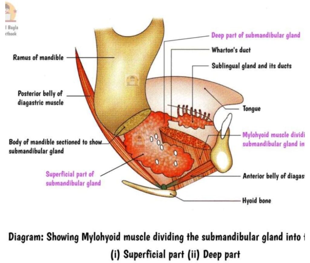
Figure 1: Mylohyoid muscle dividing the submandibular gland
Histology
Mixed tubuloacinar gland. Serous acini predominant (more than mucous), with serous demilunes of Gianuzzi capping mucous acini. Myoepithelial cells, intercalated ducts, striated ducts. More serous than sublingual, less than parotid.
Pathologies
Tumours
Only ∼8-10% of salivary tumours, but ∼50% are malignant (vs parotid 20% malignant).
Benign (50%):
Facial nerve exits stylomastoid foramen, enters gland, divides into temporofacial and cervicofacial trunks, then into 5 terminal branches at anterior border: Temporal, Zygomatic, Buccal, Marginal Mandibular, Cervical – “Two Zebras Bit My Cheeks” – forming pes anserinus. Lies between superficial and deep lobes, more superficial inferiorly.
Malignant:
- Adenoid cystic carcinoma – commonest malignant in submandibular
- Mucoepidermoid carcinoma
- Acinic cell carcinoma
- Carcinoma ex pleomorphic adenoma
- Adenocarcinoma NOS, squamous cell carcinoma
Sialolithiasis / Calculi
Commonest salivary disease. 80-90% of all salivary calculi occur in submandibular gland.
Why commoner in submandibular than parotid:
- Wharton’s duct is longer, larger caliber, tortuous, upward course against gravity
- Saliva is more alkaline, more mucinous, higher calcium and phosphate concentration – more viscous
- Flow is slower, more stagnant
- pH alkaline promotes precipitation
- Presence of mylohyoid flexure
Other:
- Cysts: ranula (plunging), mucus retention cyst, epidermoid
- Abscess: acute suppurative
sialadenitis (ascending infection)
- Neurofibroma, lipoma
- Sialadenosis
- IgG4-related disease / Kuttner tumour (chronic sclerosing sialadenitis)
Note: on Sjogren's Syndrome:
Autoimmune lymphocytic infiltration of exocrine glands. Triad: xerostomia, xerophthalmia, parotid enlargement. Submandibular involved in 50%. Histology: focal lymphocytic sialadenitis with epimyoepithelial islands. Associated with anti-Ro (SSA), anti-La (SSB), Rheumatoid factor. Risk of MALT lymphoma 44x.
Investigations
-
Plain X-ray:
Lateral oblique, AP mandible - 80% calculi radio-opaque in submandibular (vs 40% parotid) - Occlusal X-ray: For distal duct stones
-
Sialography:
Iodinated contrast into Wharton's duct - filling defects, strictures. Contraindicated in acute infection
-
Ultrasound:
First line, 90% sensitive for stones >2mm, distinguishes solid/cystic, vascularity on Doppler
-
Magnetic resonance imaging (MRI):
Gold standard for soft tissue, tumour extent, perineural spread, deep lobe, marrow invasion. MR sialography non-invasive
-
Others:
Fine needle aspiration cytology (FNAC), sialendoscopy, scintigraphy
Gold standard for tumours: MRI with gadolinium; for calculi: Ultrasound + non-contrast CT.
Management of Pathologies
-
Sialadenitis:
Hydration, sialogogues, massage, antibiotics (anti-staph), incision drainage if abscess
-
Sialolithiasis:
Conservative, sialendoscopy + basket retrieval, intracorporeal lithotripsy, transoral duct slitting, gland excision for proximal/hilar large stones with chronic sialadenitis
-
Benign tumours:
Excision of gland (with tumour) - extracapsular dissection not safe as in parotid
-
Malignant:
Gland excision + neck dissection (levels I-III) + adjuvant radiotherapy +/- chemotherapy
Step-By-Step Excision Of Submandibular Gland
Indications: Chronic sialadenitis with failed conservative, recurrent sialolithiasis (hilar/proximal), benign/malignant tumours, Kuttner tumour, ranula plunging.
Rule of 2s:
- 2 fingerbreadths below mandible
- 2 cm from angle
- 2 layers: platysma and investing fascia
- 2 arteries: facial and lingual (ligate)
- 2 nerves to preserve: marginal mandibular, hypoglossal; 1 nerve to see but preserve - lingual
- 2 veins: facial and submental
- Drain for 2 days
Procedure:
-
1. Anaesthesia:
General anaesthesia with nasotracheal or orotracheal tube away from field. Supine, neck extended, head turned opposite, shoulder roll. Hypotensive anaesthesia helpful.
-
2. Skin incision:
5-6cm curvilinear submandibular (Risdon) incision, 2 fingerbreadths (2-3cm) below and parallel to inferior border of mandible, from angle to near hyoid. Mark.
3. Elevation of flap with preservation of marginal mandibular nerve:
- Incise skin, subcutaneous tissue, platysma.
- Elevate superior flap in subplatysmal plane up to mandible.
- Identify and ligate anterior facial vein – nerve lies deep/superficial to vein. Hayes Martin: elevate vein with flap to protect nerve.
- Incise investing fascia over gland after nerve identified.
4. Dissection of superficial lobe:
- Retract gland inferiorly, identify facial artery at posterior border, ligate doubly – proximal and distal (artery grooves gland and may be encountered twice).
- Dissect capsule off mylohyoid anteriorly and digastric inferiorly. Preserve submental vessels if possible.
5. Deep lobe dissection and identification of structures:
- Retract mylohyoid anteriorly with Langenbeck. Deep lobe appears.
- Identify:
1. Lingual nerve – crosses duct from lateral to medial, has 2-3 fibres from submandibular ganglion hanging below it.
2. Wharton’s duct – below lingual nerve.
3. Hypoglossal nerve – deeper, on hyoglossus muscle, with vena comitans. - Dissect duct anteriorly, ligate and divide. Free lingual nerve.
- Identify hypoglossal, preserve.
- Ligate small vessels to lingual and facial arteries.
-
6. Haemostasis:
Bipolar cautery, check lingual, facial, submental arteries. Saline wash.
-
7. Drain:
Insert 10-14F suction drain (Redivac) through separate stab, fix.
-
8. Closure:
In layers - mylohyoid/digastric area not closed tight, platysma with Vicryl 3-0, skin subcuticular.
Complications
- Marginal mandibular nerve injury (most common 5-10%) – lip deviation
- Lingual nerve injury – loss of taste anterior 2/3, tongue anesthesia
- Hypoglossal nerve injury – tongue deviation to operated side
- Bleeding / hematoma from facial artery
- Infection, salivary fistula, sialocele
- Scar hypertrophy, keloid
- Residual stone in duct stump
- Frey syndrome – rare in submandibular excision
Management: Most nerve praxias recover 3-6 months. Physiotherapy, eye care if facial weakness, exploration if hematoma.
Methods of Managing Calculi
- Conservative: hydration, lemon drops, gland massage
- Transoral sialolithotomy for distal duct stones
- Sialendoscopy with Dormia basket for <5mm
- Laser lithotripsy (Holmium:YAG), extracorporeal shock wave
- Submandibular gland excision for hilar, intraparenchymal >10mm, recurrent infection
Note on HIV infection of Parotid
HIV-associated salivary gland disease – benign lymphoepithelial cysts, diffuse infiltrative lymphocytosis syndrome (DILS). Bilateral parotid > submandibular enlargement, CD8 infiltration, associated with cervical lymphadenopathy. Manage with HAART, aspiration, sclerotherapy, superficial parotidectomy if cosmetic.
In conclusion,
Submandibular gland though small, has complex anatomy and high propensity for calculi and malignancy compared to parotid. Understanding of fascial layers and nerve relations is key to safe excision.
Share Post On:
Recent Posts
Categories
RELATED POSTS

Get in Touch
Read doctor-produced health and medical information written for you to make informed decisions about your health concerns.
Address
Adenta
info@grovehealth.net
Working Hours
Mon - Friday @ 8am - 5 pm