
Overview of the Nasal Septum
The nasal septum is the midline partition that divides the nasal cavity into two fossae. It is critical for laminar airflow, olfaction, humidification, and support of the external nose. Pathology of the septum is one of the most common causes of nasal obstruction worldwide.
What is the Nasal Septum?
This is a composite osteo-cartilaginous structure covered on both sides by mucoperichondrium / mucoperiosteum, which provides its blood supply and separates the right and left nasal cavities from the nares anteriorly to the choanae posteriorly.
Parts of the Septum
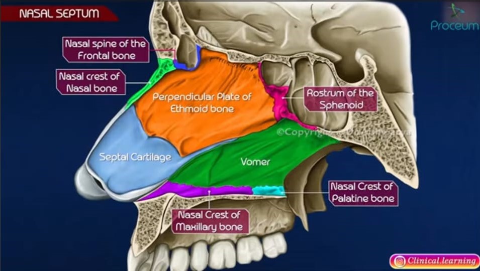
Figure 1: The parts of the Nasal Septum
-
Cartilaginous part:
Quadrangular cartilage / septal cartilage. Main anterior part. Contributes to nasal dorsum support. Joins nasal bones and medical crus of upper lateral cartilages at the keystone area.
-
Bony part:
Vomer: posteroinferior. Forms the posterior-inferior septum.
Perpendicular plate of ethmoid: posterosuperior. Thin, continues with cribriform plate superiorly.
- Nasal crest of the maxillary bone
- Nasal crest of the Palatine bone
- Nasal crest of the frontal bone.
-
Rostrum of the sphenoid
-
Fibro-fatty / Membranous part:
Columellar septum. Anterior-most small portion between the medial crura of alar cartilages and the quadrangular cartilage. Made of fibrofatty tissue and skin. Very mobile.
-
Coverings:
Septal cartilage has perichondrium; bony parts have periosteum. Both are fused with respiratory mucosa (pseudostratified ciliated columnar epithelium with goblet cells). Rich vascular plexus, especially in Little's area anteriorly.
-
Blood supply
Kiesselbach's plexus anteriorly - anastomosis of sphenopalatine, greater palatine, superior labial, anterior ethmoidal arteries. Woodruff's plexus posteriorly.
-
Innervation:
Anterior ethmoidal nerve, nasopalatine nerve, branches of greater palatine.
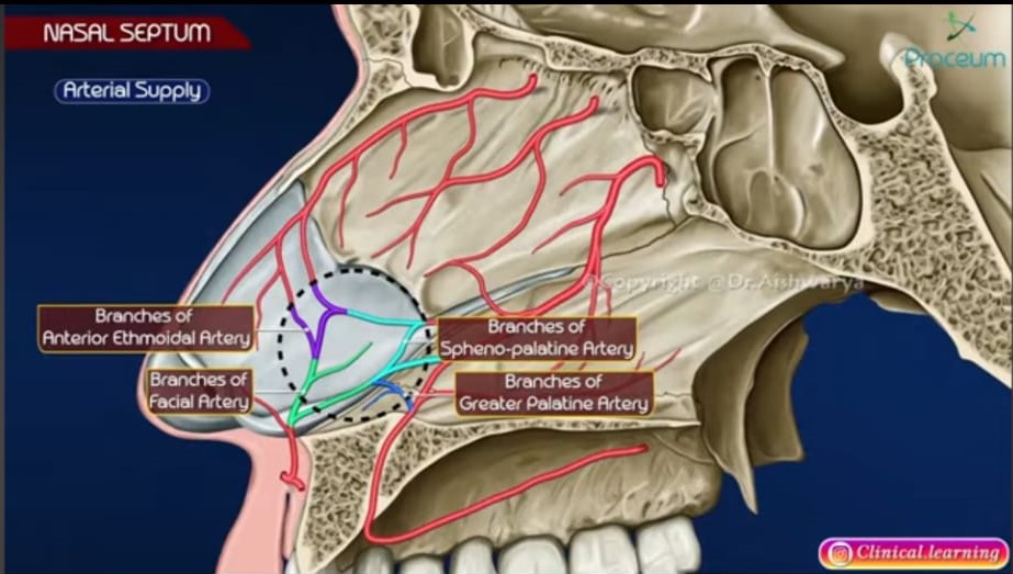
Figure 1: The Diagram of the arterial supply of the nasal septum.
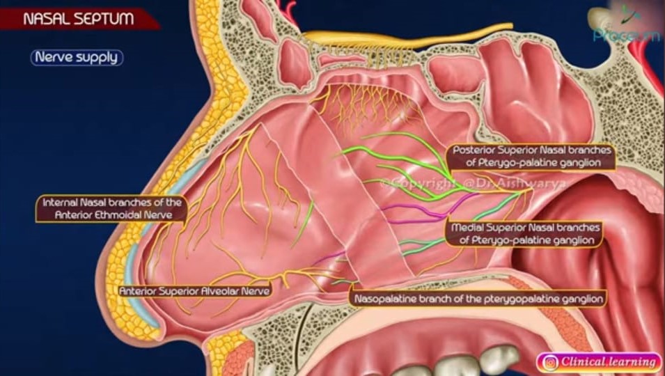
Figure 1: The Diagram of the nerve supply of the nasal septum
Epidemiology
-
Septal deviation:
Found in up to 70-80% of adults to some degree, but only ∼25-30% symptomatic. More common in males due to trauma.
-
Deviated septum due to birth trauma:
5-10% of newborns.
-
Septal perforation:
Rare; incidence <1% after septal surgery. More common in males and industrial workers.
-
Septal hematoma:
Common after nasal trauma, especially in children, due to loose mucoperichondrium.
Pathophysiology of Septal Pathology
-
Deviated Septum (DNS)
Developmental: unequal growth between septal cartilage and bony framework, pressure from surrounding structures like a high-arched palate.
-
Traumatic
Direct injury displaces cartilage off the vomerine groove. Results in C-shaped, S-shaped, spur, or caudal dislocation. Causes turbulent airflow, mucosal drying, and compensatory inferior turbinate hypertrophy on the contralateral side.
-
Septal Spur
Sharp angulation at the junction of bone and cartilage due to trauma or overgrowth. Traumatises the lateral wall.
-
Septal Perforation
Loss of cartilage/bone + mucoperichondrium on both sides. Caused by; iatrogenic (septal surgery, repeated cautery, nasal packing), trauma (nose picking), chronic inflammation (granulomatosis with polyangiitis, sarcoidosis, TB, SLE), cocaine abuse, chronic topical steroids/decongestants, tumours. Leads to loss of laminar flow and whistling.
-
Epistaxis related to septum
90% of nosebleeds are anterior from Little's area. Drying, crusting over spur/deviation, excessive picking, loss of perichondrium causes vessel exposure.
-
Septal Hematoma
Trauma ruptures submucoperichondrial vessels. Blood collects between cartilage and perichondrium. Cartilage depends on perichondrium for nutrition, so it undergoes avascular necrosis within 24-72 hours. Bacterial colonisation leads to septal abscess.
-
Septal Abscess
Infected hematoma secondary to furuncle, ethmoid sinusitis, or dental infection. Staphylococcus aureus is common. Can spread to cavernous sinus via valveless veins, cause meningitis, and cause saddle nose deformity due to cartilage destruction.
Symptoms and Signs of Septal Pathology
Symptoms
Unilateral/bilateral nasal obstruction, mouth breathing, hyposmia, headache (contact point headache), recurrent epistaxis, crusting, whistling sound on breathing in perforation, anosmia, recurrent sinusitis, snoring, cosmetic deformity.
Signs
External deviation; caudal dislocation into the vestibule; spur touching the inferior turbinate; dilated vessels in Little’s area; perforation seen as a hole on transillumination; fluctuant bluish swelling bilaterally for hematoma; tender red swelling with fever for abscess; saddle nose.
Management
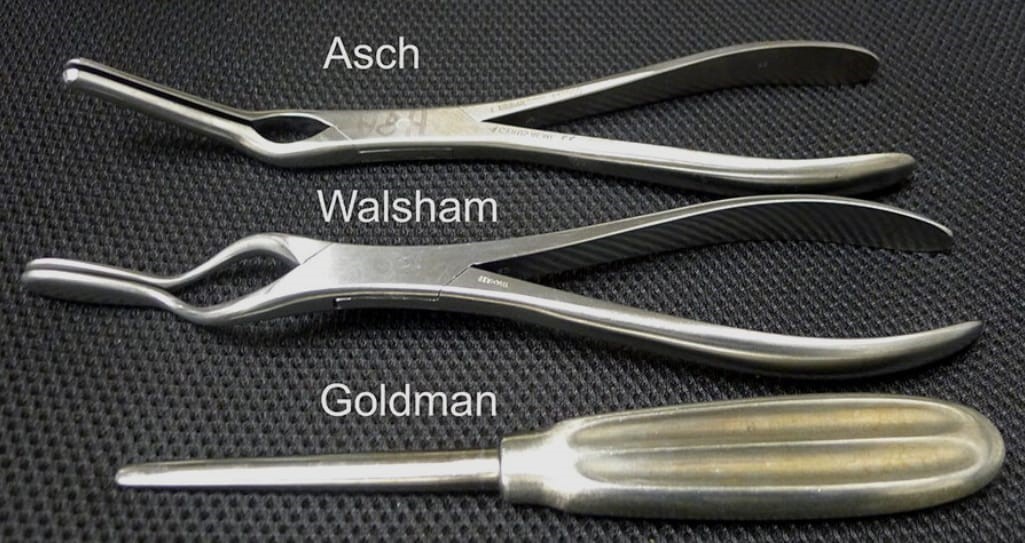
Figure 1: The Instruments for Reduction.
Clinical Notes on Septal Deviation and Use of Asch's and Walsham's Forceps
Septal deviation following trauma can be associated with fracture of the nasal bones and septum. Two types of reduction:
Asch's forceps:
Two flat blades with a slight bayonet curve. Used for septal deviation reduction. Works on cartilaginous septum. After nasal fracture, the septum is grasped between the blades and mobilised back to midline with controlled force. Often used after Walsham’s for nasal bones.
Walsham's forceps:
One blade has a rubber guard; the other is angled. Right and left pair. Used for reduction of fractured nasal bones. The guarded blade goes inside the nasal cavity to protect mucosa, outer blade on skin. Nasal bones are elevated and pushed medially/laterally.
Procedure in principle:
Under general anaesthesia or local with sedation + topical 4% lidocaine + adrenaline. Decongest nose. Assess with speculum. Walsham’s first to disimpact and elevate nasal bones, then Asch’s to straighten septum. Check patency; place nasal packs or internal splints.
Time of reduction: Crucial
- Best within a few hours before oedema sets in.
- Optimal window: within 7-10 days in adults, within 3-5 days in children because healing is faster.
- After 2 weeks: bones start to unite with callus and closed reduction fails; requires formal septorhinoplasty after 3-6 months.
- Septal hematoma is an emergency: immediate drainage within hours to prevent abscess and necrosis.
-
Indication for septoplasty vs immediate reduction:
A simple traumatic deviation without severe comminution or septal cartilage fracture can be reduced. Complex, old deviation, or obstructive spur needs elective septoplasty later.
In Conclusion,
The nasal septum is more than a partition; it is a functional and structural support for the nose. Its pathologies, especially deviation, are common and often underdiagnosed. Understanding anatomy helps differentiate benign obstruction from dangerous complications like hematoma/abscess, which need urgent drainage, and perforation, which needs systemic workup. Early recognition and timely reduction within 10 days using Asch’s and Walsham’s forceps can prevent long-term functional and aesthetic deformity.
Know more of
Septal deviation refers to a deviation or displacement of the nasal septum from the midline, which can cause nasal obstruction and other symptoms.
Share Post On:
Recent Posts
Categories
Get in Touch
Read doctor-produced health and medical information written for you to make informed decisions about your health concerns.
Address
Adenta
info@grovehealth.net
Working Hours
Mon - Friday @ 8am - 5 pm