
Comprehensive Overview of Choanal Atresia
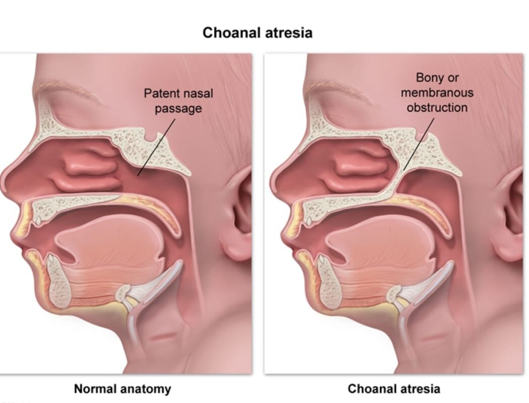
Overview
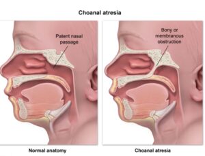
Choanal atresia is a congenital condition where the posterior nasal aperture is blocked. It’s one of the more common congenital anomalies of the nose and is an important cause of neonatal respiratory distress because newborns are obligate nasal breathers for the first few months of life.
Choanal atresia
Is the complete or partial obstruction of the choana, the opening between the nasal cavity and nasopharynx. It can be unilateral or bilateral, and the obstruction can be bony, membranous, or mixed.
Epidemiology
1. Incidence: ∼1 in 5,000 to 8,000 live births
2. Sex ratio: More common in females, ∼2:1 female to male ratio
3. Laterality: ∼60% unilateral, 40% bilateral
3. Side for unilateral: Slightly more common on the right side
Embryology
Normally, the nasal and oral cavities are separated during weeks 4-6 of gestation. The nasobuccal membrane and oronasal membrane break down to form the choana around week 7.
The leading theory for atresia: persistence of the buccopharyngeal membrane, or failure of recanalization of the nasal fossa after the nasal plugs resolve at ∼10 weeks. There may also be abnormal mesoderm migration to the choanal region.
Pathophysiology
Newborns breathe primarily through the nose for the first 4 – 6 weeks.
Bilateral atresia :
Causes acute airway obstruction immediately after birth. The infant can breathe while crying but desaturates and becomes cyanotic when the mouth is closed and quiet.
Unilateral atresia :
Often asymptomatic in newborns because the other side allows nasal breathing. It may present later with chronic unilateral rhinorrhea, nasal obstruction, or recurrent sinusitis.
Pathology and CHARGE Associations
Pathology: Histologically, bony atresia shows thick bone separating the nasal cavity from nasopharynx. Membranous type is a thin fibrous membrane. Mixed type has both.
CHARGE Association: Choanal atresia is a major criterion for CHARGE syndrome.
- CHARGE = Coloboma, Heart defects, Atresia choanae, Retardation of growth/development, Genital hypoplasia, Ear anomalies
- ∼ 50 – 60% of patients with bilateral choanal atresia have CHARGE syndrome
- Overall, 30 – 50% of all choanal atresia cases are associated with other anomalies.
Percentage of association with other anomalies:
1. CHARGE: 10-20% of all choanal atresia cases
1. Crouzon, Pfeiffer, Treacher Collins: Craniofacial syndromes
2. Cardiac defects: 10-15%
3. TCNS anomalies: 5-10%
4. Tracheoesophageal fistula: Seen in VACTERL association
All bilateral cases should be screened for associated anomalies.
Types of Choanal Atresia
By tissue:
1. Bony: ∼ 90% of cases. Thick bony plate, usually at vomer
2. Membranous: ∼ 10% of cases. Thin mucosal membrane
3. Mixed : Combination of bone and membrane
By laterality:
1. Unilateral: ∼ 60% of cases
2. Bilateral: ∼ 40% of cases. This is the life-threatening form in newborns
Symptoms and Signs
Bilateral:
- Cyclic cyanosis: cyanotic at rest, pink when crying
- Respiratory distress immediately after birth
- Inability to pass a 6Fr catheter through both nostrils to nasopharynx
- Relief of distress with an oral airway
Unilateral:
- Chronic unilateral foul rhinorrhea
- Nasal obstruction on one side
- Often diagnosed later in infancy or childhood
- Failure to pass catheter on one side only
Investigations
-
Clinical test:
Inability to pass catheter 5-6 cm into nasopharynx
-
CT scan of paranasal sinuses with bone window:
Gold standard. Defines thickness of atresia plate, type bony vs membranous, and vomer width. Done after stabilization.
-
Nasal endoscopy:
To visualize the atretic plate
-
Screening for associated anomalies:
Echocardiogram, renal US, ophthalmology exam, hearing test if CHARGE suspected
-
Genetic testing:
For CHD7 gene if CHARGE suspected
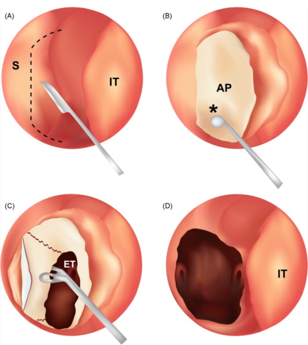
Figure 1
Endoscopic choanoplasty
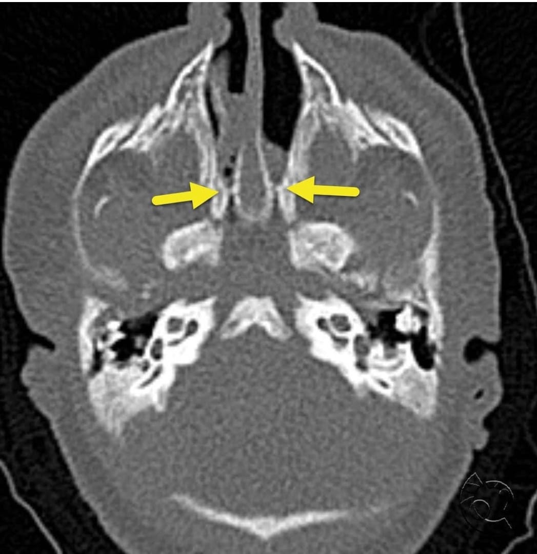
Figure 2
Axial CT Scan of PARANASAL
Management
Emergency Management - mainly for bilateral cases
- Place an oral airway to bypass the obstruction
- Intubation if needed
- Position infant prone to keep mouth open
- Stabilize before any definitive workup
Definitive Management
Treatment is surgical repair. Timing depends on presentation and laterality.
Proposed age for definitive surgical intervention:
-
Bilateral:
Emergency in neonatal period, ideally within first few days to weeks of life once stable.
-
Unilateral:
Can be delayed. Traditionally 6 months to 2 years to allow facial growth, but many centers now do it earlier, 3-6 months, to improve nasal breathing and development
Success statistics by age:
Data varies by center and technique. Overall success rates:
- Neonatal repair: 60 – 70% primary success. Higher revision rate due to smaller anatomy and granulation.
- Infant repair 3 – 12 months: 70 – 85% success.
- After 1 year: 80 – 90% success. Lower restenosis rates.
Revision rates overall are 20-40%, mostly due to restenosis.
Methods of Surgical Intervention
-
1. Transnasal endoscopic repair:
Current standard. Drill or microdebrider to open atretic plate, remove bone, create neochoana. Minimally invasive.
-
2. Transpalatal approach:
Open approach through palate. Used for very thick bony atresia or revision cases. More morbidity.
-
3. Transseptal approach:
Less common now.
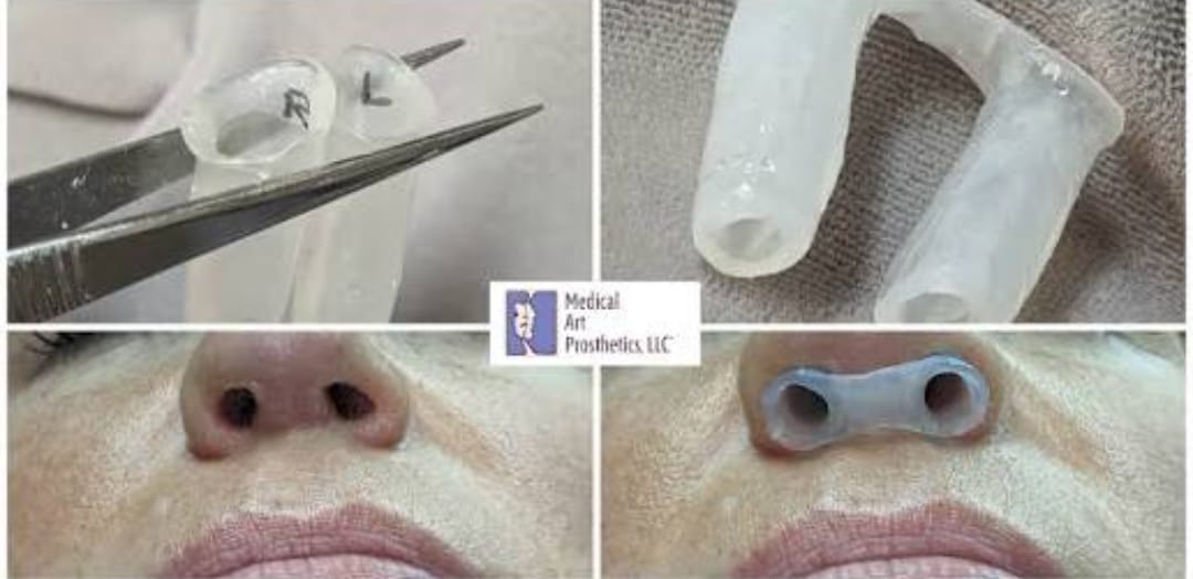
Figure 3
Customized nasal stents for Choanal atresia
Stent vs No Stent:
-
With stent:
Silicone stent placed for 4-6 weeks post-op. Aims to prevent restenosis. Higher rates of granulation, infection, and patient discomfort. Still widely used, especially in neonates.
-
No stent:
Growing evidence that stents may not improve outcomes and may increase complications. Some surgeons do stentless repair with mitomycin C and aggressive post-op debridement.
Current trend: Selective stenting. Bilateral cases and revision cases more likely to get stents.
Complications of Surgery
- Restenosis: Most common, 20-40%
- Granulation tissue formation
- Synechiae
- CSF leak: Rare, if skull base violated
- Hemorrhage
- Septal perforation
- Velopharyngeal insufficiency: More with transpalatal approach
- Need for revision surgery
Conclusion and Note on Surgery and Timing
Choanal atresia is a potentially life-threatening congenital anomaly, especially when bilateral. Early recognition in the newborn period is critical. While emergency airway management is the first priority, definitive repair timing is tailored: urgent in bilateral cases for survival, and more elective in unilateral cases to balance surgical success with facial growth.
Endoscopic transnasal repair is now the preferred method with good success rates, but restenosis remains the main challenge. The role of stents is evolving, with increasing support for stentless techniques in selected patients. Because of the high association with CHARGE and other syndromes, a multidisciplinary evaluation is essential for all patients.
Share Post On:
Recent Posts
Categories
Get in Touch
Read doctor-produced health and medical information written for you to make informed decisions about your health concerns.
Address
Adenta
info@grovehealth.net
Working Hours
Mon - Friday @ 8am - 5 pm