Comprehensive Overview of Adenoids & Adenoid Facies

Overview
The adenoids are part of Waldeyer’s ring — the body’s first line of immune defense in the upper airway. In children, they’re physiologically large and shrink after puberty. Chronic hypertrophy leads to mouth breathing, sleep issues, and the classic facial changes called “adenoid facies”.
What is Adenoids?
Adenoids is the nasopharyngeal tonsil. A collection of lymphoid tissue in the roof and posterior wall of the nasopharynx.
What is Adenoid facies?
Adenoid facies is the constellation of craniofacial, dental, and facial soft-tissue changes resulting from long-term nasopharyngeal obstruction and obligate mouth breathing during growth.
Anatomy & Histology - Nasopharynx & Position
-
Location:
Midline, on the roof and posterior wall of the nasopharynx, just behind the choanae and above the soft palate.
-
Histology:
Non-encapsulated lymphoid tissue covered by pseudostratified ciliated columnar respiratory epithelium with goblet cells. Contains lymphoid follicles with germinal centers, similar to other mucosa-associated lymphoid tissue [MALT].
Figure 1
Location of adenoids.
Blood Supply
Arterial supply: Primarily the ascending pharyngeal artery branch of external carotid. Also contributions from the ascending palatine, pharyngeal branch of maxillary, and sphenopalatine arteries.
Venous drainage: Via the pharyngeal venous plexus and internal jugular vein.
Lymphatic Drainage
Drains to the retropharyngeal nodes, then to the deep cervical chain, especially the upper jugulodigastric nodes. This is why nasopharyngeal infections can present with upper neck nodes.
Contraindications for adenoidectomy
1. Cleft palate or submucus palate
2. Acute upper respiratory infection
3. Bleeding order
4. Other medical problem where surgery or anesthesia is contraindicated.
Innervation
1. Sensory: Maxillary nerve V2 via the pharyngeal branch and sphenopalatine ganglion.
2. Autonomic: Parasympathetic from the sphenopalatine ganglion.
Nasopharyngeal Tonsils (adenoids) vs Palatine Tonsil
| Adenoids | Tonsils | |
|---|---|---|
|
Location |
Roof / posterior nasopharynx, midline. |
Oropharynx, between palatoglossal & palatopharyngeal arches |
|
Capsule |
No true capsule |
Encapsulated |
|
Crypts |
No deep crypts |
Multiple deep crypts |
|
Surface Epithelium |
Respiratory epithelium |
Non-keratinized stratified squamous |
|
Peak Size |
3 – 6 years old |
6 – 10 years old |
Development of Adenoids
Present at birth. Arise from lymphoid tissue aggregation in the nasopharyngeal submucosa. Become prominent after exposure to antigens in infancy.
Growth Pattern of Adenoids:
Birth – 3y: Rapid growth
3 – 6y: Peak size, often largest relative to nasopharyngeal airway
After puberty: Physiological involution/atrophy due to hormonal changes. Rarely symptomatic in adults unless pathologic.
Investigations
1. Plain X – ray of soft tissues of nasopharynx in lateral view.
2. Digital palpitation of adenoid with index fingure.
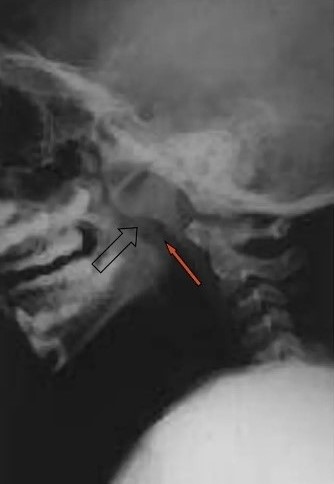
Figure 2
X-ray of the nasopharynx
Physiology of Adenoids
Part of the mucosal immune system. Trap inhaled antigens, produce IgA, and initiate immune responses. They are not essential after early childhood because other lymphoid tissue compensates.
Bacteria & Viruses That Infect Adenoids
Viruses: Adenovirus, RSV, rhinovirus, influenza, EBV
Bacteria: 2Streptococcus pneumoniae, Haemophilus influenzae, Moraxella catarrhalis, Group A
Streptococcus: These form biofilms and can be a reservoir for chronic rhinosinusitis or otitis media.
Enlarged Adenoids
Aetiology of Enlarged Adenoids
Recurrent infection / inflammation: Repeated viral/bacterial URIs
Allergy: Allergic rhinitis and chronic mucosal edema
Genetic / familial: Tendency to lymphoid hyperplasia
Environmental: Smoke exposure, pollution
Clinical Features of Enlarged Adenoids
Nasal: Chronic nasal obstruction, mouth breathing, hyponasal / “blocked” voice, rhinorrhea, snoring, OSA
Aural: Eustachian tube obstruction and otitis media with effusion, conductive hearing loss, “glue ear”
Craniofacial: See “adenoid facies” below
Adenoidectomy
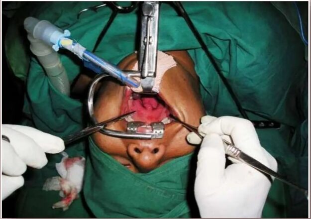
Figure 3
Adenoidectomy
Adenoidectomy may be indicated alone or combined with tonsillectomy. Adenoids are removed first and nasopharynx packed before starting tonsillectomy.
Adenoid Facies: Features & Explanation
This develops over years of chronic nasal obstruction during facial growth. It’s a clinical sign, not a diagnosis.
-
1. Dull / Blank Look
Why: Chronic fatigue from poor sleep + OSA, plus open-mouth posture and reduced facial muscle tone. The face looks “expressionless”.
-
2. Pinch Nose / Narrow Nostrils
Why: Lack of normal nasal airflow reduces stimulus for alar cartilage and nasal base growth. Mouth breathing bypasses the nose.
-
3. Open Mouth
Why: Compensatory mouth breathing. Lips cannot stay closed at rest because nasal airflow is blocked.
-
4. Crowded Upper Teeth
Why: Tongue rests low due to open mouth. Without tongue pressure against the palate, transverse maxillary growth is reduced and narrow arch and crowding.
-
5. High Arched Palate / “Gothic Palate”
Why: Tongue not up against the palate to widen it. The palate grows vertically instead of transversely.
-
6. Receded Upper Lip
Why: Constant open mouth posture and lack of lip seal. Upper lip becomes hypotonic and short.
-
7. Receded Chin / Retrognathic Mandible
Why: Downward and backward mandibular rotation to open the airway. Reduced anterior facial height growth.
-
Maxillary Hypoplasia
Why: Reduced nasomaxillary complex growth due to lack of nasal breathing stimulus. Contributes to midface retrusion.
-
9. Elongated Face / “Long Face Syndrome”
Why: Increased lower facial height from mandibular rotation + vertical maxillary growth. Overall dolichofacial pattern.
-
10. Dennie-Morgan Lines
Why: Extra folds of skin under the lower eyelid. Classically associated with atopy/allergy, which commonly coexists with adenoid hypertrophy.
-
11. Allergic Shiners
Why: Venous congestion under the eyes from chronic nasal/allergic inflammation. Gives dark bluish-purple periorbital discoloration.
-
12. Allergic Salute
Why: Habitual upward rubbing of the nose with the palm to relieve itching/congestion. Over time causes a transverse nasal crease.
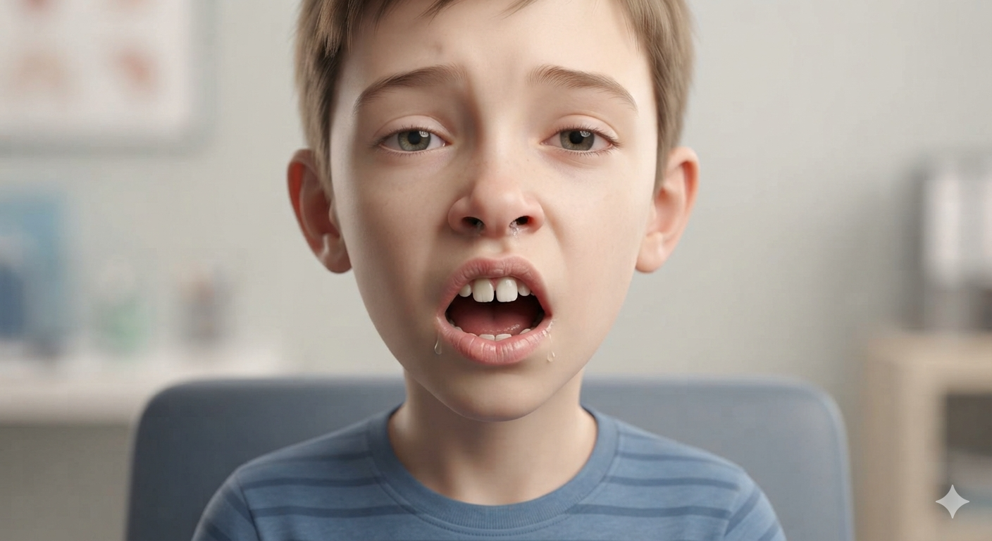
Figure 4
Adenoid Facies – Child, Frontal View
To conclude,
Many children have 1 – 2 features without adenoid issues.
Diagnosis requires ENT exam, nasal endoscopy, or lateral cephalogram / X-ray + correlation with symptoms.
Adenoids Summary
- Definition
Definition: Nasopharyngeal tonsil. Lymphoid tissue on roof/posterior wall of nasopharynx. Part of Waldeyer’s ring.
- Anatomy / Histology
Midline nasopharynx. Covered by respiratory epithelium. Non-encapsulated with lymphoid follicles.
- Blood Supply
Arterial: Ascending pharyngeal, ascending palatine, sphenopalatine.
Venous: Pharyngeal plexus IJV.
- Lymphatic Drainage
Retropharyngeal nodes deep cervical chain.
- Innervation
Sensory V2 via sphenopalatine ganglion. Parasympathetic from same.
Development & Physiology
Development:
- Present at birth, grow with antigen exposure.
Growth Pattern:
- Rapid 0-3y, peak 3-6y, involutes after puberty
Physiology:
- Mucosal immunity, IgA production. Not essential later in life.
Aetiology of Hypertrophy:
- Recurrent infection, allergy, genetics, smoke/pollution.
Viruses:
- Adenovirus, RSV, EBV.
Bacteria:
- S. pneumoniae, H. influenzae, M. catarrhalis, GAS.
Clinical Features of Hypertrophy
-
Nasal:
Obstruction, mouth breathing, hyponasal voice, snoring, OSA
-
Aural:
Eustachian tube block OME, conductive hearing loss
-
Craniofacial:
Adenoid facies
Adenoid Facies: Features & Why
Management: Adenoidectomy + bilateral grommet insertion. At 6 weeks: TM clear, tympanometry Type A, speech therapy started. Hearing normalized.
Key point: Address adenoids in < 4yr olds.
-
Dull / Blank Look
Poor sleep + OSA fatigue, reduced facial tone
-
Pinch Nose / Narrow Nostrils
Lack of nasal airflow during growth
-
Open Mouth
Chronic mouth breathing to bypass obstruction
-
Crowded Upper Teeth
Low tongue posture narrow maxillary arch
-
High Arched Palate
Tongue not widening palate during growth
-
Recessed Upper Lip
Hypotonic lip from open-mouth posture
-
Recessed Chin
Mandibular rotation backward to open airway
-
Maxillary Hypoplasia
Reduced nasomaxillary growth without nasal breathing
-
Elongated Face
Increased lower facial height, dolichofacial pattern
-
Dennie-Morgan Lines
Extra fold under eye, linked to atopy/allergy
-
Allergic Shiners
Venous congestion from chronic nasal inflammation
-
Allergic Salute
Upward nose rub and transverse crease on bridge of the nose.
Clinical Note: Medical trial 6 – 12 weeks is common before surgery, unless severe OSA, growth failure, or recurrent infection are present.
Share Post On:
Recent Posts
Categories
Get in Touch
Read doctor-produced health and medical information written for you to make informed decisions about your health concerns.
Address
Adenta
info@grovehealth.net
Working Hours
Mon - Friday @ 8am - 5 pm