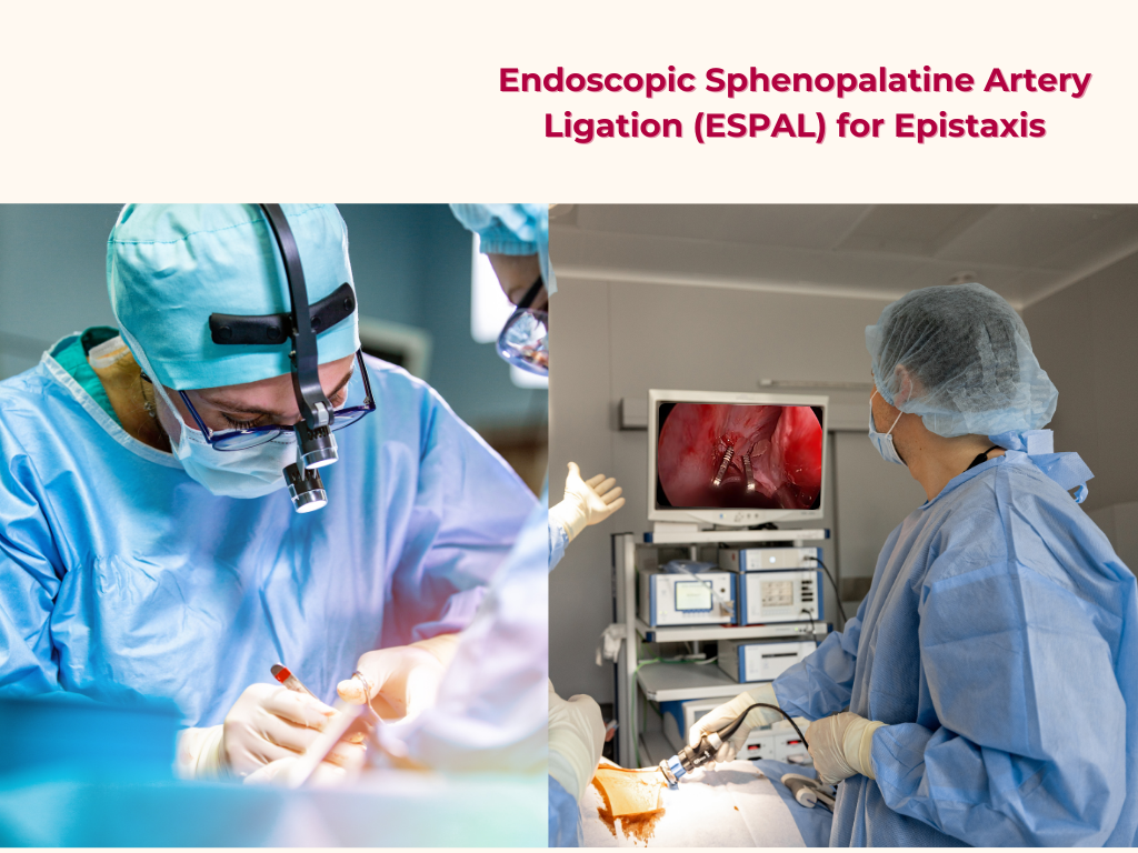
Endoscopic Sphenopalatine Artery Ligation (ESPAL) for Epistaxis

Overview
Endoscopic Sphenopalatine Artery Ligation (ESPAL) is a minimally invasive surgical procedure used to control severe or recurrent epistaxis (nosebleed) by ligating the sphenopalatine artery, which is a common source of nasal bleeding.
Anatomy of the Sphenopalatine Artery
The sphenopalatine artery is a branch of the maxillary artery that supplies blood to the nasal cavity. It is a crucial artery in the nasal cavity and plays a significant role in the management of epistaxis (nosebleed).
Origin
The sphenopalatine artery arises from the maxillary artery in the pterygopalatine fossa.
Course
The artery passes through the sphenopalatine foramen, which is located in the posterior aspect of the lateral nasal wall.
Branches
The sphenopalatine artery gives off several branches that supply the nasal cavity, including
Distribution
The sphenopalatine artery supplies blood to the posterior parts of the nasal cavity, including the lateral wall and the nasal septum.
Importance
The sphenopalatine artery is a common source of bleeding in epistaxis, particularly in the posterior nasal cavity. Ligation or cauterization of this artery may be necessary to control severe or recurrent bleeding.
Clinical Significance
Understanding the anatomy of the sphenopalatine artery is essential for:
-
1. Managing epistaxis
Knowledge of the artery's location and branches is crucial for effective treatment.
-
2. Nasal surgery
Surgeons need to be aware of the artery's location to avoid injury during procedures.
In summary, the sphenopalatine artery is a vital artery in the nasal cavity that plays a significant role in supplying blood to the posterior parts of the nose. Its anatomy is essential for understanding and managing nasal bleeding and other nasal conditions.
Indications for ligation
-
1. Severe or recurrent epistaxis
When conservative measures fail to control bleeding.
-
2. Failed nasal packing or cauterization
When other treatments are ineffective.
Preoperative Preparation
-
1. Antibiotics
May be administered to prevent infection.
-
2. Tranexamic acid
May be used to reduce bleeding.
-
3. Blood transfusion
May be necessary if significant blood loss has occurred.
Operative Process
Anesthesia
The procedure is typically performed under general anesthesia.
Position of Patient
The patient is positioned in a supine position with the head elevated.
Types of Endoscope
-
0-degree endoscope
Provides a direct view of the nasal cavity.
-
30-degree endoscope
Offers a wider field of view and is useful for visualizing the sphenopalatine artery.
Procedure
-
1. Uncinectomy
Removal of the uncinate process to access the posterior fontanelle.
-
2. Middle meatal antrostomy
Creation of an opening in the middle meatus to access the sphenopalatine foramen.
-
3. Localization of the sphenopalatine artery
Identification of the artery and its branches.
-
4. Ligation of the main artery and branches
Clipping or cauterization of the sphenopalatine artery and its branches.
Management
Postoperative Management
-
1. Nasal packing
May be used to control bleeding and support the nasal mucosa.
-
2. Monitoring
Close monitoring of the patient's vital signs and nasal bleeding.
-
3. Nasal irrigation
Saline irrigation may be used to keep the nasal cavity clean and promote healing.
-
4. Intranasal corticosteroids
May be used to reduce inflammation and promote healing.
-
5. Liquid paraffin
May be applied to the nasal cavity to prevent crusting and promote healing.
Microscopy Compared to Endoscopy in SPAL
Endoscopy is generally preferred over microscopy for ESPAL due to its ability to provide a wider field of view and better illumination.
Contraindications to Endoscopic SPAL
-
1. Severe nasal septal deviation
May make it difficult to access the sphenopalatine artery.
-
2. Nasal tumors or masses
May obstruct the view or make it difficult to access the artery.
-
3. Previous nasal surgery
May make the procedure more challenging due to altered anatomy.
Endoscopic sphenopalatine artery ligation is a effective and minimally invasive procedure for controlling severe or recurrent epistaxis. Proper preoperative preparation, surgical technique, and postoperative management are essential for optimal outcomes.
Want to Know More of
Nosebleeds (epistaxis) can be alarming, but most cases can be managed with simple first aid. Here’s what you need to know. . . . . . . . . . . . . . . . . . . . .
Share Post On:
Recent Posts
-
Nuggets of ORL-OTOLOGY
-
Nuggets of ORL-RHINOLOGY
-
Nuggets of Otorhinolaryngology-Basic sciences
-
Anatomy of the Muscles of the Soft Palate
-
Ethmoidal Arteries Ligation for Epistaxis
-
Submucous Cleft Palate (SMCP)
-
Approach to Ligation of the External Carotid Artery
-
Approach to Managing a 3-Year-Old Boy with a Foreign Body in the nasal cavity.
-
Approach to Managing a 3-Year-Old Boy with a Foreign Body impacted in the ear canal.
-
Endoscopic Sphenopalatine Artery Ligation (ESPAL) for Epistaxis
-
Surgical Management of Epistaxis
-
Technique of Incision and Drainage of Septal Hematoma/Septal Abscess
-
Upper Aerodigestive Tract Foreign Body Impaction
-
Incision and Drainage of Hematoma Auris
-
Rigid Bronchoscopy for Retrieval of Foreign Bodies in Children
-
Foreign Body Impaction in the Larynx, Trachea, and Bronchi
-
Leadership Position is a Tool, not a Trophy
-
Carcinoma of the Oropharynx
-
Peritonsillar Abscess
-
Ethics of Doctor-Patient Relationship
-
Doctor-Patient Relationship Case Scenarios
-
Asymmetrical Tonsils and Approach to Evaluation and Management
-
Nasal Polyposis
-
Rigid Oesophagoscopy and Complication
-
Anatomy of Oesophagus
-
Stridor, Snoring, Stertor And Wheezing: How They Compare
-
Temporomandibular Joint (TMJ)
-
Otoacoustic Emissions
-
Tympanometry
-
Functional Endoscopic Sinus Surgery (FESS)
-
Tracheostomy
-
Clinical Voice Test (CVT) for Hearing Loss
-
Acute Epiglottitis And Approach To Management
-
Synoptic Overview Of Nasopharyngeal Carcinoma
-
Prioritizing Support For People With Disabilities Over Unhealthy Competitions That Marginalise The Downtrodden
-
Otitic Barotrauma
-
Titbits of Informed Consent Process for a Medical or Surgical Procedure
-
Comprehensive Overview of Mpox (Monkeypox)
-
Overview Of Corrosive Ingestion - Acid & Alkalis, and Management Approach
-
Ethical Conundrum
Categories
RELATED POSTS
Get in Touch
Read doctor-produced health and medical information written for you to make informed decisions about your health concerns.

