Submucous Cleft Palate (SMCP)

Overview
Cleft palate is a congenital anomaly that affects the roof of the mouth, occurring when the tissues that form the palate do not fuse properly during fetal development. Cleft palate can occur in isolation or as part of a broader syndrome. Submucous cleft palate (SMCP) is a subtype of cleft palate, characterized by a defect in the musculature of the soft palate, despite an intact mucosal surface.
What is Submucous Cleft Palate?
Submucous cleft palate is a congenital anomaly where there is a defect in the muscles of the soft palate, but the overlying mucosa is intact. This can lead to velopharyngeal insufficiency (VPI), affecting speech and feeding.
Epidemiology
The prevalence of submucous cleft palate is estimated to be around 1 in 1,200 to 1 in 2,000 live births. However, the true prevalence may be higher due to underdiagnosis.
Embryology, Pathology, and Pathophysiology
During embryonic development, the palate forms from the fusion of the medial nasal prominence and the maxillary prominence. In submucous cleft palate, the muscles of the soft palate (tensor veli palatini, levator veli palatini, and musculus uvulae) fail to fuse properly, leading to a deficiency in the velopharyngeal sphincter.
Degrees or Severity and Classification of Submucous Cleft
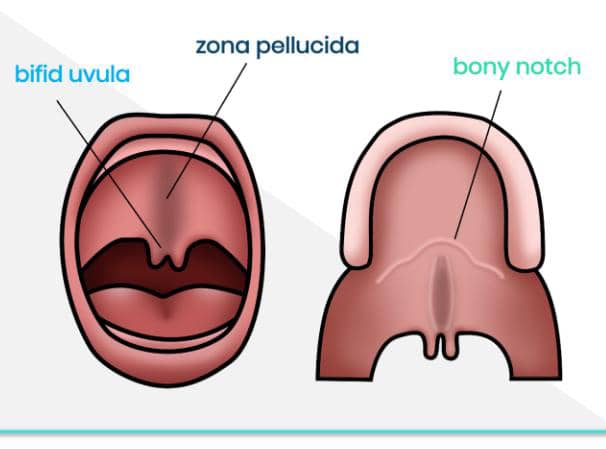
Submucous cleft palate can be classified based on the severity of the muscular defect and the presence of symptoms. The Calnan criteria are often used to diagnose SMCP, which include:
- Bifid uvula
- Zona pellucida (a translucent area in the midline of the soft palate)
- Muscular diastasis (a separation of the muscles in the soft palate)
Clinical Association
Association with Maternal Age at Conception
Advanced maternal age has been associated with an increased risk of cleft palate, including submucous cleft palate.
Association with Down's Syndrome, Pierre Robin's, and Treacher Collins Syndrome
Submucous cleft palate is more common in individuals with certain genetic syndromes, including:
-
1. Down's syndrome
Individuals with Down's syndrome are at higher risk of having a submucous cleft palate.
-
2. Pierre Robin sequence
This condition is characterized by micrognathia, glossoptosis, and often, a cleft palate or submucous cleft palate.
-
3. Treacher Collins syndrome
This genetic disorder affects the development of bones and other tissues of the face, and may include cleft palate or submucous cleft palate.
Symptoms and signs
Symptoms of submucous cleft palate may include:
-
1. Speech difficulties
Hypernasal speech, nasal emission, or articulation errors.
-
2. Feeding difficulties
Infants may experience feeding problems, such as nasal regurgitation.
-
3. Recurrent ear infections
Due to Eustachian tube dysfunction.
Diagnosis
Features at Physical Examination
The physical examination may reveal:
- Bifid uvula
- Notching of the hard palate
- Zona pellucida
- Muscular diastasis
Investigations to Confirm Diagnosis
Diagnosis can be confirmed through:
-
1. Nasopharyngoscopy
Visualizing the velopharyngeal sphincter during speech.
-
2. Videofluoroscopy
Assessing velopharyngeal function during speech.
-
3. Imaging studies
Such as MRI or CT scans, may be used in some cases.
Complications of Submucous Cleft
Untreated submucous cleft palate can lead to:
1. Speech difficulties: Persistent speech problems.
2. Hearing problems: Recurrent ear infections or hearing loss.
3. Feeding difficulties: Persistent feeding problems.
Management of Submucous Cleft
Management typically involves a multidisciplinary team, including:
-
1. Speech therapy
To address speech difficulties.
-
2. Surgical intervention
To repair the muscular defect and improve velopharyngeal function.
Age for Surgical Intervention
The timing of surgical intervention varies depending on the severity of symptoms and the child's overall health. Some surgeons recommend repair between 6 months to 2 years of age.
Submucous cleft palate is a congenital anomaly that requires early diagnosis and management to prevent long-term complications. A multidisciplinary team approach is essential for optimal outcomes.
Share Post On:
Recent Posts
-
Nuggets of ORL-OTOLOGY
-
Nuggets of ORL-RHINOLOGY
-
Nuggets of Otorhinolaryngology-Basic sciences
-
Anatomy of the Muscles of the Soft Palate
-
Ethmoidal Arteries Ligation for Epistaxis
-
Submucous Cleft Palate (SMCP)
-
Approach to Ligation of the External Carotid Artery
-
Approach to Managing a 3-Year-Old Boy with a Foreign Body in the nasal cavity.
-
Approach to Managing a 3-Year-Old Boy with a Foreign Body impacted in the ear canal.
-
Endoscopic Sphenopalatine Artery Ligation (ESPAL) for Epistaxis
-
Surgical Management of Epistaxis
-
Technique of Incision and Drainage of Septal Hematoma/Septal Abscess
-
Upper Aerodigestive Tract Foreign Body Impaction
-
Incision and Drainage of Hematoma Auris
-
Rigid Bronchoscopy for Retrieval of Foreign Bodies in Children
-
Foreign Body Impaction in the Larynx, Trachea, and Bronchi
-
Leadership Position is a Tool, not a Trophy
-
Carcinoma of the Oropharynx
-
Peritonsillar Abscess
-
Ethics of Doctor-Patient Relationship
-
Doctor-Patient Relationship Case Scenarios
-
Asymmetrical Tonsils and Approach to Evaluation and Management
-
Nasal Polyposis
-
Rigid Oesophagoscopy and Complication
-
Anatomy of Oesophagus
-
Stridor, Snoring, Stertor And Wheezing: How They Compare
-
Temporomandibular Joint (TMJ)
-
Otoacoustic Emissions
-
Tympanometry
-
Functional Endoscopic Sinus Surgery (FESS)
-
Tracheostomy
-
Clinical Voice Test (CVT) for Hearing Loss
-
Acute Epiglottitis And Approach To Management
-
Synoptic Overview Of Nasopharyngeal Carcinoma
-
Prioritizing Support For People With Disabilities Over Unhealthy Competitions That Marginalise The Downtrodden
-
Otitic Barotrauma
-
Titbits of Informed Consent Process for a Medical or Surgical Procedure
-
Comprehensive Overview of Mpox (Monkeypox)
-
Overview Of Corrosive Ingestion - Acid & Alkalis, and Management Approach
-
Ethical Conundrum
Categories
RELATED POSTS
Get in Touch
Read doctor-produced health and medical information written for you to make informed decisions about your health concerns.

