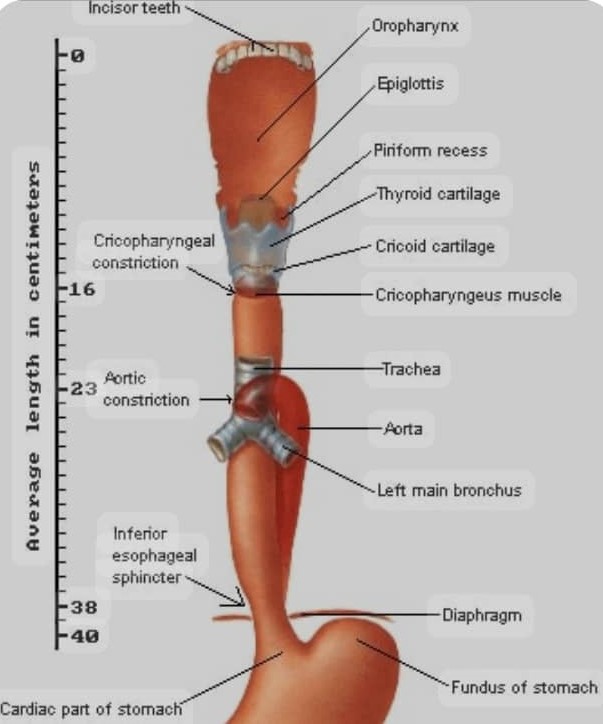
Overview
Rigid oesophagoscopy is a medical procedure that involves the insertion of a rigid tube with a light and camera (oesophagoscope) into the oesophagus to visualize, diagnose, and treat various oesophageal conditions.
Rigid oesophagoscopy has been used for decades to manage oesophageal disorders, particularly those requiring intervention, such as foreign body removal or dilation of strictures.
The development of rigid oesophagoscopy dates back to the early 20th century, with significant advancements in instrumentation and technique over the years.
Indications for Rigid Oesophagoscopy
Diagnostic
1. Evaluation of dysphagia odynophagia, or unexplained chest pain.
2. Biopsies of oesophageal tumours
Therapeutic
1. Removal of foreign bodies such as impacted coins, dentures, fish bones, meat bolus etc.
2. Dilatation of strictures.
3. Treatment of bleeding lesions etc.
Types
-
Adults oesophagoscopes:
Various lengths and diameters available.
-
Paediatric oesophagoscopes:
Smaller sizes for use in children.
Adult Rigid Oesophagoscopes
Types
-
1. Standard rigid oesophagoscopes:
These are the most commonly used type, available in various lengths and diameters.
-
2. Rigid oesophagoscopes with distal lighting:
These scopes have a light source at the distal end, providing better illumination of the oesophageal lumen.
-
3. Rigid oesophagoscopes with suction channels:
These scopes have a suction channel that allows for simultaneous suctioning and visualization.
Sizes
1. Diameter: Adult rigid oesophagoscopes typically range from 5-14 mm in diameter.
2. Length: The length of adult rigid oesophagoscopes can vary from 20-50 cm.
Common Sizes
1. Small: 5-7 mm diameter, 20-30 cm length
2. Medium: 8-10 mm diameter, 30-40 cm length
3. Large: 11-14 mm diameter, 40-50 cm length.
Paediatric Rigid Oesophagoscopes
Types
-
1. Standard pediatric rigid oesophagoscopes:
These scopes are designed for general use in pediatric patients.
-
2. Pediatric rigid oesophagoscopes with distal lighting:
These scopes provide better illumination of the oesophageal lumen.
-
3.Pediatric rigid oesophagoscopes with suction channels:
These scopes allow for simultaneous suctioning and visualization.
Sizes
1. Diameter: Pediatric rigid oesophagoscopes typically range from 3-7 mm in diameter.
2. Length: The length of pediatric rigid oesophagoscopes can vary from 15-30 cm.
Common Sizes
1. Neonatal: 3-4 mm diameter, 15-20 cm length
2. Infant: 4-5 mm diameter, 20-25 cm length
3. Pediatric: 5-7 mm diameter, 25-30 cm length.
The choice of rigid oesophagoscope size and type depends on the specific procedure, patient anatomy, and the surgeon’s preference.
Selection of Size of Oesophagoscope:
The size of the oesophagoscope is selected based on the patient's age, size, and the specific procedure being performed.
Step-by-Step Procedure of Rigid Oesophagoscopy
General anaesthesia or sedation is typically used.
The oesophagoscope is introduced into the oropharynx under direct vision.
The scope is advanced through the cricopharyngeal sphincter with gentle pressure.
The scope is advanced carefully, visualizing the oesophageal lumen.
The scope is withdrawn slowly, inspecting the oesophageal mucosa.
Post-Operative Monitoring
Patients are monitored for complications, such as bleeding, perforation, or respiratory distress.
Complications of Oesophagoscopy
1. Perforation: A serious complication that requires prompt management.
2. Bleeding: May occur during or after the procedure.
3. Respiratory complications: Aspiration or airway obstruction.
Detecting Esophageal Perforation
-
1. Clinical signs:
Severe chest pain, fever, tachycardia, or subcutaneous emphysema.
-
2. Imaging:
Chest X-ray or CT scan may show signs of perforation, such as pneumomediastinum or pleural effusion.
Management of Oesophageal Perforation
-
1. Immediate action:
Stop the procedure, and assess the patient's condition.
-
2. Conservative management:
May be considered for small, contained perforations.
-
3. Surgical intervention:
Often required for larger or non-contained perforations.
-
4. Antibiotics:
Broad-spectrum antibiotics are typically administered.
-
5. Supportive care:
Patients may require intensive care unit (ICU) admission and supportive therapy.
Prompt recognition and management of oesophageal perforation are crucial to prevent serious complications and improve patient outcomes.
Share Post On:
Recent Posts
-
Nuggets of ORL-OTOLOGY
-
Nuggets of ORL-RHINOLOGY
-
Nuggets of Otorhinolaryngology-Basic sciences
-
Anatomy of the Muscles of the Soft Palate
-
Ethmoidal Arteries Ligation for Epistaxis
-
Submucous Cleft Palate (SMCP)
-
Approach to Ligation of the External Carotid Artery
-
Approach to Managing a 3-Year-Old Boy with a Foreign Body in the nasal cavity.
-
Approach to Managing a 3-Year-Old Boy with a Foreign Body impacted in the ear canal.
-
Endoscopic Sphenopalatine Artery Ligation (ESPAL) for Epistaxis
-
Surgical Management of Epistaxis
-
Technique of Incision and Drainage of Septal Hematoma/Septal Abscess
-
Upper Aerodigestive Tract Foreign Body Impaction
-
Incision and Drainage of Hematoma Auris
-
Rigid Bronchoscopy for Retrieval of Foreign Bodies in Children
-
Foreign Body Impaction in the Larynx, Trachea, and Bronchi
-
Leadership Position is a Tool, not a Trophy
-
Carcinoma of the Oropharynx
-
Peritonsillar Abscess
-
Ethics of Doctor-Patient Relationship
-
Doctor-Patient Relationship Case Scenarios
-
Asymmetrical Tonsils and Approach to Evaluation and Management
-
Nasal Polyposis
-
Rigid Oesophagoscopy and Complication
-
Anatomy of Oesophagus
-
Stridor, Snoring, Stertor And Wheezing: How They Compare
-
Temporomandibular Joint (TMJ)
-
Otoacoustic Emissions
-
Tympanometry
-
Functional Endoscopic Sinus Surgery (FESS)
Categories
RELATED POSTS
Get in Touch
Read doctor-produced health and medical information written for you to make informed decisions about your health concerns.

