
Questions & Answers
Nuggets of ORL-Head & Neck Surgery
More To Read
41. What is a myocutaneous flap? Give examples used in head and neck surgery.
Answer:
A myocutaneous flap is a flap which includes muscle, fascia, subcutaneous tissue, skin and feeding blood vessels in one unit. It is axial in design and contains a major blood vessel which runs along the undersurface providing perforating vessels to supply muscles and skin. Examples used in head and neck reconstruction include :
- pectoralis major
- latissimus dorsi
- trapezius
- sternocleidomastoid
- supraclavicular flap
42. Describe the features and treatments of “Singers node’s”(vocal cord nodules).
Answer:
Singer’s nodes is a condition that usually affects professional voice users and screaming children’. Following either excessive or inappropriate use of the voice, the patient complains of hoarseness of voice and inability to reach high notes without extra effort. On indirect laryngoscopy white, symmetrical, sessile nodules are seen at the junction of the anterior third and posterior two thirds of the vocal cords. The nodules are less than 2 mm in size. Pathologically there is benign localised keratosis.
Treatments include;
- voice rest
- elimination of focal sepsis in the URT
- microlaryngoscopic excision if the problem persists despite voice rest
- speech therapy
43. What are the main parotid tumours in children?
Answer:
In children up to 60% of persistent parotid enlargement is caused by
- haemangioma,
- lymphangioma, and
- neurofibroma, where as these tumours are causes of parotid swelling in the adults.
Other causes of parotid enlargement in children are
- chronic sialdenitis,
- leukaemias, and
- lymphomas.
44. What is Behcet’s syndrome?
Answer:
This an association of
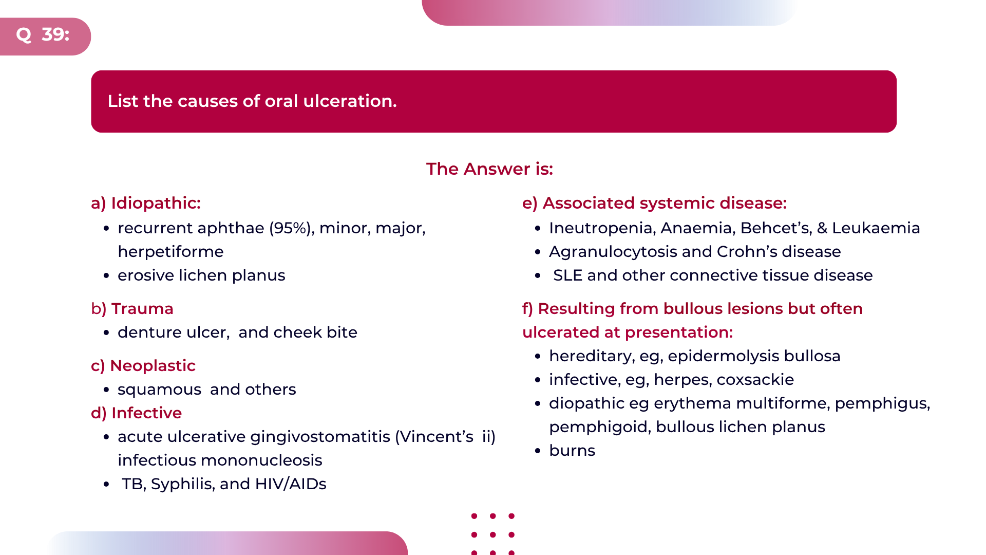
a) oropharyngeal ulceration (aphthous type)
b) genital ulceration and
c) iritis.
In this idiopathic condition multisystem involvements is now recognised. Encephalitis and blindness are rare complications.
Steroids, azathioprine and cyclophosphamide may be used in the severe case.
45. Describe the features of adenolymphoma of the parotid gland.
Answer:
This tumour has the synonyms of papillary cystadenoma lymphomatosum
and Warthin’s tumour. It accounts for 20% of the benign tumours of the parotid gland. It is a disease of the elderly and affects approximately 7 men to 1 woman.
Lymphoid element is prominent on histological examination and the tumour is believed to arise from a residue of salivary duct epithelium within an intraparotid lymph node.
As a rule, there is never malignant degeneration. Clinically, they may appear suddenly and fluctuate in size. These soft and cystic tumours are bilateral in 10% of cases.
Occasionally they are discovered as incidental findings during an operation on the neck. Treatment is usually with superficial parotidectomy to obtain a histopathological diagnosis. Fine needle aspiration biopsy may alter this treatment policy.
46. List the causes of recurrent laryngeal nerve paralysis.
Answer:
- Malignancy eg. Carcinoma of bronchus, oesophagus, thyroid and metastatic carcinoma of the nasopharynx
- Surgical trauma eg surgery to thyroid, oesophagus and great vessels
- Idiopathic
- Inflammatory eg. Apical or mediastinal scarring due to Tuberculosis
- Non-surgical trauma eg. Sharp and blunt injuries to the neck.
- Miscellaneous eg. Connective disorders (rheumatoid arthritis), syphilis, thrombosis of subclavian vein
- Stretching of the nerve eg. By an aortic aneurysm, non-malignant thyroid or parathyroid disease , an enlarged heart or lymph nodes.
- Neurological
- Peripheral Neuritis eg. Diabetes, alcoholism, lead poisoning, viral.
- central and general neurological disorders eg. Cerebrovascular accident, syringomyelia, multiple sclerosis.
47. Describe the features of a thyroglossal cyst.
Answer:
This is a cyst which develops in the line of a persistent thyroglossal duct which runs from the foramen caecum through or in front of the hyoid bone to the thyroid gland.
The thyroglossal cyst, the commonest cause of a midline cyst , usually presents in childhood.
No age group, however, is exempt. In the majority there is a painless midline mobile cyst which moves upwards on swallowing or protrusion of the tongue. Occasionally the cyst may be situated just lateral to the midline (usually left) and atypical presentation includes infection and fistulisation.
Treatment involves excision of the cyst plus the duct including the body of the hyoid bone.
48. What is the most common respiratory problem arising from glandular fever and how may the problem be managed?
Answer:
In glandular fever there may be a very prominent tonsillar enlargement such that the tonsils may even meet in the midline.
These tonsils are hard and this contribute to the symptoms of dysphagia and oral respiratory difficulty.
The managements includes:
- confirm the diagnosis
- admission to hospital if any complications develop
- most patients will breathe comfortably through the nose. Those patients with nasal obstruction may be helped with vasoconstrictor nasal drops or by insertion of nasopharyngeal airway
- steroids to reduce the tonsillar swelling
- The role of antibiotics is controversial. In the presence of a superadded infection they may be of use. Ampicillin should be avoided
- general support such as adequate hydration and oxygen if appropriate
- rarely a tracheostomy may be required.
49. Describe the features and classification of a laryngocoele.
Answer:
This is an air or fluid containing prolongation of the laryngeal saccule from the ventricle. It is believed that the saccule becomes encysted secondary to ostial obstruction. Should the cyst discharge then fluid is replaced by air in the sac. It is commoner in elderly men.
Most are unilateral and there is an association of increased incidence of laryngocoele with laryngeal carcinoma.
Laryngocoeles can be
- External- herniating around or through the thyrohyoid membrane and presenting as a neck swelling which expands on coughing and empties on pressure
- Internal- confined within the laryngeal framework
- Combined external and internal.
A muffled and hoarse voice is usually present. Radiographic examination during the valsalva manoeuvre may demonstrate an air containing lesion.
Treatment of the internal type involves marsupialising the lesion endoscopically, where as the external approach and excision is required for the external type.
50. How may the auriculo-temporal or Frey’s syndrome be managed?
Answer:
Management may include:
- Reassurance – Up to 20% of parotidectomy patients may complain of this
symptom. The majority improve within 6 months - Topical therapy – The drying agents that are available in antiperspirants may be used to good effect, such as zinc oxide
- Anticholinergics – such as atropine or hyoscine have been used but the side
effects are often worse than the condition and the results are poor - Surgical – The objective is to interrupt the parasympathetic fibres.
i) re-evaluate the skin flap over the parotid
ii) section of the auriculo-temporal nerve
iii) perform a tympanic neurectomy and chorda tympanectomy
iv) perform a middle cranial fossa geniculate ganglion and lesser petrosal nerve
section. Of these the tympanic neurecttomy is at present the favoured method. The results, however, are not as good as might be expected.
51. Describe the features of Hashimoto lymphocytic thyroiditis
Answer:
This common cause of a bilateral , often asymmetrical goitre, affects 9 females to 1 male. The gland consistency varies from firm and glandular to obviously nodular. History and examination suggest the diagnosis with confirmation by histology (usually from fine needle aspiration) which shows focal lymphocyte infiltration. There is a gross elevation of thyroid auto-antibodies. All ranges of thyroid function are seen; however, myxoedema is the usual outcome. A normal T4 and T3 with a raised TSH is nearly pathognomonic of the condition. Treatment with thyroxine usually prevents further growth of the goitre and may encourage shrinkage. There is debate as to whether there is an increased incidence of neoplasia in Hasimoto’s disease. At present this association is believed to be coincidental.
52. Describe the features of papillary carcinoma of the thyroid.
Answer:
About1 70% of thyroid carcinomas are papillary. This is the commonest childhood thyroid cancer and not an unusual incidental post mortem finding. It occurs in any age group but has a peak incidence in the third and fourth decades (20- 40 years). About 75% of patients are females. This rarely encapsulated tumour is usually infiltrating.
Histologically, there are papillary projections into a variable colloid stroma with characteristics psammoma bodies. Up to 20% of tumours may be multifocal within the thyroid gland on simple examination, and histological examination may increase this number 90%.
Presentation is usually with a solitary thyroid nodule although lymphatic metastases may be present. Investigation with ultrasound and isotope scanning may be helpful. Fine needle aspiration biopsy (FNAC) has a high diagnostic yield, with diagnostic accuracy of 94%, sensitivity of 85-95% and specificity of above 90%.
In cases of doubt surgical exploration is required. Total lobectomy, total thyroidectomy and functional neck dissection all have a place in the surgical management depending depending on the tumour extent.
Metastatic disease can be diagnosed using a radioactive iodine scan and ablated with a therapeutic dose of radio-iodine. Thyroxine is given to those patients post treatment both as replacement therapy and as TSH suppression.
NB: There is evidence that differentiated thyroid carcinoma is TSH dependent.
53. Describe the aetiology, features and managements of myasthenia gravis.
Answer:
This is an autoimmune acquired disorder which causes skeletal muscle weakness and fatigue.
Under the age of 40 more women are affected while over that age males predominate. Peak age incidence depends on the presence of a thymoma.
It is associated with other autoimmune disorders. A serum IgG antibody binds acetylcholine post-synaptic receptor at the neuromuscular junction and interferes with normal neuromuscular transmission.
The muscle weakness is increased by exercise and may be exacerbated by pregnancy, stress or infection. Specific muscle groups may be differentially affected.
Investigations include:
- anti-acetylcholinesterase2 receptor antibody,
- striated muscle antibody,
- edrophonium test,
- electromyograpy and
- anterior mediastinal radiology to identify those cases with an associated thymoma.
Treatment include:
- anticholinesterae drugs (pyridostigmine or neostigmine)
- prednisolone and azathioprine as second choice therapy
- plasma exchange in the severely ill
- thymectomy and
- tracheostomy for severe breathing difficulties.
54. Describe the features of syringomyelia
Answer:
Syringomyelia is a rare condition in which there is cavitation of the spinal cord extending over several segments. Damage occurs mostly to the anterior horn cells, spinothalamic and corticospinal fibres.
Syringobulbia is when cavitation extends into the medulla and pons. Predisposing factors include, congenital and acquired abnormalities at the foramen magnum such as basal arachnoiditis, posterior fossa tumours and intermedullary gliomas.
The usual presentation is between the ages of 10 and 30 and may be:
- with hand neurotrophic ulceration, weakness or wasting.
- dissociated sensory loss to pain and temperature in one or both arms with associated
burning shoulder pain and painless destruction of the joints of the hand. - cerivcal lesions above c5 may cause coarse rotatory nystagmus, vertigo, Horner’s
syndrome and ataxia. The earliest features of syringobulbia may be sensory loss over the face, dysphagia and dysphonia. Investigation is with myelography, CT and MRI scanning.
Treatment is by surgical decompression at the the cranio- vertebral junction plus control of any secondary hydrocephalus.
55. Define reflux oesophagitis and summarise its predisposing factors.
Answer:
There is regurgitation of gastric juice into the lower oesophagus where the vulnerable squamous epithelium is injured. The reflux may be intermittent or persistent.
Reflux is caused by:
- incompetence of the lower oesophageal sphincter which may be congenital or acquired
1) anatomical eg. Scleroderma
2) physiological eg. Cigarette smoking - increased intra abdominal pressure eg. Pregnancy
- hiatus hernia (sliding)
- obesity in which fat infiltration causes loss of the fundo-oesophageal angle
- vomiting.
56. Describe the features of motor neurone disease.
Answer:
This disease is characterised by progressive degeneration of anterior horn cells, corticospinal and motor nuclei in the medulla. Although all parts of the motor system may be involved the disease may start in any area.
The most common presentation is upper limb wasting with lower limb spasticity where as within the head and neck pseudobulbar, or progressive bulbar palsy may be a feature of the disease.
The aetiology is unknown; progression is relentless; treatment does not alter the progression; males are affected more often than females; it is generally a condition of the elderly. Survival from the time of diagnosis is on average 3 – 4 years; prevalence is 5 per 100, 000.
Presentation is often insidious with diagnosis being considered when there is muscle wasting and fasciculation without associated sensory signs. The clinical diagnosis may be corroborated by EMG and muscle biopsy.
The major aspects of treatment are supportive to cope with increasing disability. Surgery may be considered for intractable aspiration.
57. Give the T part of the TNM classification of supraglottic carcinoma of the larynx.
Answer:
AJCC 8th edition:
- Tis – carcinoma in situ. To-no evidence of primary tumour
- Tx – the minimum requirements to assess the primary tumour cannot be met
- T1a – tumour confined to the laryngeal surface of the epiglottis or to an aryepiglottic fold or to a ventricular cavity or to a ventricular band.
- T1b – tumour involving the epiglottis and extending to the ventricular cavities or bands.
- T2 – tumour with extension to adjacent sites or to the glottis without fixation.
- T3 – tumour confined to the larynx but with fixation of cords and/or other evidence of deep invasion.
- T4 – tumour extended outside the larynx.
58. List the causes of macroglossia
Answer:
- Lymphatic obstruction to the tongue due to submandibular space disease
- Haemangioma
- Myxoedema
- Acromegaly
- Amyloid
- Cysts
- Actinomycosis
- Von Gierke’s disease
- Down’s syndrome
- Tumours of the tongue
- Angioneurotic oedema
- Pierre Robin ( relative macroglossia).
59. Describe the features of pseudobulbar palsy.
Answer:
This is a spastic paralysis which selectively involves the bulbar motor nuclei. The most common cause is a cerebral vascular accident. Other possibilities include;
- Multiple sclerosis (MS),
- Neoplasia, and
- Motor Neurone Disease (MND).
There is degeneration of the pyramidal fibres high in the brain stem. Owing to the rich bilateral innervation of the cranial nerve nuclei by the upper motor neurones from each cerebral hemisphere, a unilateral lesion of the pyramidal tract causes few and transitory signs within the cranial nerve territory (with the exception of the facial muscles).
In a bilateral lesion there is often a near complete paralysis of the muscles used in chewing, swallowing, and speaking. The effects are similar to bulbar palsy but without wasting or fasciculation.
- The tongue is slow moving.
- Speech is slurred and monotonous.
- The face is immobile and expressionless although facial emotional responses may be excessive, uncontrollable and inappropriate.
- The jaw jerk is increased.
- Gait may be tottery and shuffling.
- Inhalation pneumonia is the most frequent cause of death.
There is no curative treatment for this condition although in some cases surgery to protect the lower respiratory tract may be appropriate, e.g. Cricopharyngeal myotomy, Epiglottopexy or even Laryngectomy.
60. How would you confirm the diagnosis and investigate a suspected carotid body tumour in a fit 40 year-old man?
Answer:
1. Carotid angiography would be the investigation of choice.
Characteristic features are that of an egg-shell like mass displacing the internal carotid artery laterally and widening the bifurcation between internal and external carotid arteries
2. Urinary VMA to identify
- secreting carotid body tumours and
- associated secreting paragangliomas such as phaeochromocytoma
61. Define obstructive sleep apnoea.
Answer:
Sleep apnoea is characterized by repetitive episodes of upper airway tract obstruction during sleep. There are at least 30 apnoeic episodes in both REM and non-REM sleep during 7 hours of un-sedated sleep.
In the following;
- the premature infant, apnoea > 20 seconds,
- an infant, apnoea > 15 seconds and
- an adult, apnoea > 10 seconds are considered pathological
62. List the causes of sleep apnoea.
Answer:
1. Peripheral / obstructive:
- enlarged tonsils and adenoids
- nasal obstruction (deviated nasal septum, nasal pack)
- nasopharyngeal abnormalities (stenosis or incompetence)
- Pharyngeal obstruction
i) lingual tonsil, vallecular cyst
ii) abnormal deposits in the walls (obesity, myxoedema, acromegaly) - laryngeal obstruction ( webs, polyps, cysts)
- craniofacial abnormalities (Treacher collin’s , Pierre Robin)
2. Central
- high altitude
- encephalitis
- brain stem disease
- ondine’s curse
- myxoedema
- supratentorial masses
- congestive cardiac failure
3. Some cases have a combined peripheral and central aetiology.
63. List the complications associated with acute tonsillitis.
Answer:
1. Local:
- Chronic tonsillitis
- Quinsy
- Parapharyngeal abscess
- Suppurative cervical adenitis
- Acute suppurative otitis media
2. Systemic; particularly related Group A beta-haemolytic streptococcus: .
- Rheumatic fever
- Acute Glomerulonephritis
- Chorea
- Subacute bacterial endocarditis
64. What aetiological factors are associated with malignancy of the oral cavity?
Answer:
- Smoking, particularly reverse smoking of cigar or cigarette. Chewing tobacco and betel nut. Pipe smoking is associated with lower lip squamous carcinoma
- Spirits
- Spices
- Syphilis
- Spikes (sharp tooth); ill-fitting dentures and sharp teeth causing irritation
- Sunlight , particularly related to carcinoma of the lip
- Avitaminosis
- Plummer – Vinson syndrome
65. What is the site incidence of carcinoma of the tongue?
Answer:
- Lateral border – 85%
- Midline – 5%
- Tip – 5%
- Within – 5%
66. What forms of treatment may be used for a patient with intractable aspiration?
Answer:
- Nasogastric tube: This is a temporary solution and nutrition may be supplied through the tube. The problems of inhalation of saliva and complications of the tube occur
- Cuffed tracheostomy or nasotracheal tube is useful in the short term but not the long term due to complications.
- Polytetrafluoroethylene (PTFE) cord injection. There is a place for this method in high vagal paralysis but it is not very effective.
- Cricopharyngeal myotomy
- Bilateral chorda tympani and tympanic nerve sections or radiotherapy to reduce saliva
- Gastrostomy
- Separate larynx and trachea by closure at the first tracheal ring and creation of a tracheostomy
- Epiglottopexy
- The vocal cords are sutured together and a tracheostomy created
- Tracheo-oesophageal anastomosis and tracheostomys
- Total laryngectomy
67. List the advantages and disadvantages of an abscess tonsillectomy performed for quinsy.
Answer:
Advantages:
- Avoids stab incision to drain Quinsy
- Avoids interval tonsillectomy
- Allows drainage of loculated pus behind the lower pole
- Fewer attachments between tonsil and capsule
Disadvantages:
- Rupture of the abscess and inhalation occurring during the induction of anaesthesia
- Excessive haemorrhage from contralateral tonsillectomy
- Infection may be spread in the opened tissue planes.
68. What are the clinical features of hysterical aphonia?
Answer:
This functional paralysis of the laryngeal adductors during phonation occurs most frequently in emotionally unstable individuals particularly in young women.
The aphonia is sometimes complete but more often the voice is reduced to a whisper. Onset and recovery are usually sudden.
The characteristic signs on indirect laryngoscopy are, failure of the cords to meet on attempted phonation, with a normal cough at which the glottis is seen to close. .
69. Summarise the treatment of atrophic laryngitis.
Answer:
a). Control associated conditions of the nose and pharynx.
b). Control irritative factors such as associated sinus disease, smoke and dusty atmospheres.
c). Loosen and remove crusts by
- inhalation of steam or menthol
- laryngeal sprays such as 8.4% sodium bicarbonate or
benzdyamine hydrochloride 0.15%
70. How do you investigate a patient with suspected sleep apnoea syndrome?
Answer:
a). CXR and ECG for signs of Cor pulmonale
b). Polysomnography which may include:
- ENG recording of eye movements
- EEG
- Strain gauges to chest and diaphragm
- A throat microphone to record airflow
- Continuous ECG for dysrhythmias
- Transcutaneous PO₂ monitorings :
71. Describe the features and treatments of a thyroid crisis.
Answer:
This rare life-threatening exacerbation of hyperthyroidism occurs in the inadequately prepared and toxic patient during the post- operative phase of thyroid surgery. The rare crisis can be encountered in the untreated thyrotoxic stressed by infection or unrelated surgery.
The features include:
- distress,
- tachycardia,
- dyspnoea,
- pyrexia,
- confusion ,
- vomiting and
- diarrhoea.
72. Classify thyroid neoplasia.
Answer:
Benign:
- Adenoma: microfollicular, foetal, hurtle cell, papillary
Malignant
- Differentiated: papillary carcinoma, follicular carcinoma, squamous carcinoma
- Undifferentiated: anaplastic
- Malignant lymphoma
- Medullary carcinoma
- Secondary
73. What methods are available to treat achalasia?
Answer:
- Medical treatment with antispasmodics eg. amylnitrite, gives poor results
- Self-dilatation with Hurst mercury bougies
- Surgical dilatation with;
Negus hydrostatic bag, Starck expanding metal dilators, Plummer bags - Heller’s cardiomyotomy
- Resection of the lower end of the oesophagus and anastomosis
74. What is the association and clinical significance of osteomas of the face and skull with colonic polyps?
Answer:
This is Gardner’s syndrome, an autosomal dominant condition comprising
- multiple sebaceous cysts,
- osteomas of the face and skull,
- dermoid tumours,
- multiple fibromas and
- colonic polyps.
The polyps are premalignant and the finding of such a patient requires that the rest of the family should also be investigated.
75. Describe the features of papillary carcinoma of the thyroid.
Answer:
About 70% of thyroid carcinomas are papillary. This is the commonest childhood thyroid cancer and not an unusual incidental post mortem finding.
It occurs in any age group but has a peak incidence in the third and fourth decades (20- 40 years). About 75% of patients are females. This rarely encapsulated tumour is usually infiltrating.
Histologically, there are papillary projections into a variable colloid stroma with characteristics psammoma bodies.
Up to 20% of tumours may be multifocal within the thyroid gland on simple examination, and histological examination may increase this number 90%.
Presentation is usually with a solitary thyroid nodule although lymphatic metastases may be present.
Investigation with ultrasound and isotope scanning may be helpful. Fine needle aspiration biopsy (FNAC) has a high diagnostic yield, with diagnostic accuracy of 94%, sensitivity of 85-95% and specificity of above 90%.
In cases of doubt surgical exploration is required. Total lobectomy, total thyroidectomy and functional neck dissection all have a place in the surgical management depending depending on the tumour extent.
Metastatic disease can be diagnosed using a radioactive iodine scan and ablated with a therapeutic dose of radio-iodine. Thyroxine is given to those patients post treatment both as replacement therapy and as TSH suppression.
NB: There is evidence that differentiated thyroid carcinoma is TSH dependent.
76. Outline the Bethesda System for Reporting Thyroid
Answer:
The system categorizes FNAC results into six categories, helping to standardize reporting and guide clinical management.
- Category I: non-diagnostic or unsatisfactory
- Category II: Benign
- Category III: Atypia of undetermined significance or follicular lesion of undetermined significance
- Category IV: Follicular neoplasm or suspicious for follicular neoplasm
- Category V: suspicious for malignancy
- Category VI: Malignant
77. Describe the features and treatment of port wine stain.
Answer:
This congenital pigmented, macules, usually of the face is caused by multiple abnormal superficial blood vessels (mainly venules) which lie immediately deep to the surface of the skin.
Surgical excision is only advisable for the very small lesions as excision and grafting may result in a far uglier deformity. The application of facial cosmetics gives good results. Laser treatment is being evaluated.
78. Classify the causes for a white patch on the oral mucosa.
Answer:
- Developmental
- Trauma / irritation
- Tobacco
- Burns (chemical or physical)
- Infection: candida, syphilis, Tuberculosis
- Dermatoses: lichen planus, discoid SLE, psoriasis, Keratosis follicularis (Darier-white’s disease)
- Neoplasia: papilloma, carcinoma in situ, carcinoma, others
- Miscellaneous: Leukoplakia, Fordyce spots, Vit A. deficiency, skin grafts.
79. Describe the course of a 2nd arch (cervicopharyngeal ) fistula
Answer:
The internal opening is usually in the supratonsillar fossa or in the upper half of the posterior faucial pillar, either anteriorly , posteriorly or on its free edge. The tract runs inferiorly passing between the internal and external carotid arteries. It courses superficial to the glossopharyngeal and hypoglossal nerves but deep to the posterior belly of the diagastric. It opens at the anterior border of the sternocleidomastoid, usually at the junction of the upper third (1/3) and lower two-thirds (2/3).
80. In which group of patients are you most likely to see a cold abscess in the neck and what are the stages?
Answer:
The group of likely patients include:
- Immunosuppressed, like HIV/ AIDS
- Diabetics
- Elderly
- Malnourished
- People with chronic illnesses
- No-caucasian and non-mongolian races. The Irish ???
The stages of cold abscess in the neck are:
- Stage 1: lymph node enlargement- firm, painless swelling of lymph nodes
- Stage 2: caseation – Nodes become softer, fluctuant
- Stage 3: collar-stud abscess- pus collects under the deep cervical fascia, may point to the skin.
- Stage 4: sinus formation- Abscess ruptures through the skin, forming a draining sinus.
81. Following a glottic biopsy the pathology is reported as a carcinoma insitu. What is the subsequent management?
Answer:
Most surgeons would advise and perform micro-laryngoscopic stripping of the affected vocal cord to ensure that all disease has been fully excised and that there is not more invasive process which has not been biopsied. Assuming the subsequent specimen shows no invasive disease then regular follow-up is required.
82. What are the symptoms and investigations, that you will use to confirm pharyngeal pouch?
Answer:
The symptoms of pharyngeal pouch are:
- Dysphagia, often long-standing
- Regurgitation of undigested food and
- Halitosis. This discomfort may be caused by distension of the pouch. The patient takes longer and longer to complete a meal. He may complain of a swelling in the neck on the left side which may gurgle and empty on external pressure.
- Cough, due to overspill, and
- Weight loss are other common features.
The investigations include:
- Barium swallow study: This is the most common test, where you swallow a contrast medium (barium) to outline the pouch on x-rays.
- Endoscopy (oesophago-gastro-duodenoscopy): This visualises the pouch and rule out other conditions like cancer.
- Manometry: measures pressure in the oesophagus, assessing sphincter function.
83. What is the significance of pooling of saliva above cricopharyngeus as seen on indirect laryngoscopy?
Answer:
This is Jackson’s sign and is associated with severe dysphagia or aphagia. It is due to obstruction caused by
- Intrinsic, mural or extrinsic mass or foreign body
- Neuromuscular incoordination of any cause, eg. Motor neurone disease.
- Acute painful inflammatory or infective conditions of the oesophagus or pharynx eg. Candidiasis
84. Summarise the treatment of acute spontaneous rupture of the oesophagus.
Answer:
- Resuscitation
- Gastrograffin diagnostic swallow
- Thoracotomy with insertion mediastinal and pleural drains. Primary repair only if the oesophageal wall will hold the sutures. This usually is possible only within the first 24 hours
- Adequate appropriate systemic antibiotics
- Good nutritional support preferably through a feeding jejunostomy.
85. Classify laryngitis
Answer:
Laryngitis is divided into Acute and Chronic.
ACUTE:
- Infective: Bacterial and Viral
- Non-infective: Voice misuse, Trauma, Chemical I, Thermal, Radiotherapy and Allergy.
CHRONIC:
- Infective: TB, syphilis, lupus, leprosy, scleroma, Fungal
- Non-infective (specific):
Sarcoid, Wegener’s, Rheumatoid arthritis, Sleroderma, Pemphigus, Amyloid, Laryngeal joint arthropathies. - Non-infective (Non-specific):
i) without hyperplasia
ii) with hyperplasia .eg. contact pachydermia, interarytenoid pachydermia and circumscribed epithelial hyperplasia - Non-classified: vocal polyps, polypoid degeneration, dysphonia plicae ventricularis I, vocal nodules, atrophic conditions
86. Classify benign laryngeal tumours
Answer:
- Epithelia tumours: papilloma, single or multiple juvenile
- Connective tissue tumours: fibroma, chondroma, angioma, lipoma, rhabdomyoma, leiomyoma, granular cell myoblastoma
- Non-specific: intubation granuloma, salivary tumours in ectopic tissue, amyloid, polypoid, degeneration, laryngeal cysts
87. Outline the treatments of acute laryngitis.
Answer:
Treatment include:
- Voice rest
- Steam inhalations
- Soothing sprays, lozenges
- Apply warmth to the neck
- Cough linctus
- Rest
- Avoidance of smokey, dusty atmospheres, alcohol, and smoking
- Systemic antibiotics if infection is present
88. Give the aetiological factors associated with chronic laryngitis.
Answer:
- Faulty voice production
- Infection in teeth, tonsils or sinuses
- Excess use of alcohol or tobacco
- Dust or irritant fumes
- Incomplete resolution of acute laryngitis
- Mouth breathing
89. Describe the management of a patient who has received a direct blow to the larynx.
Answer:
Immediate security of the compromised airway is paramount. Apply advanced ATLS protocol.
History:
Suspicion of an injury is crucial Dyspnoea, dysphagia and pain may be present
Examination:
- External palpation to identify surgical emphysema and loss of the normal neck contour. Loss of laryngeal crepitus may indicate a retropharyngeal haematoma
- IDL and nasendoscopy: This may identify oedamtous haemorrhagic arytenoids, mucosal tears and disorganised vocal cords. The epiglottis may be detached.
Investigations:
Radiography can identify laryngeal swelling, emphysema and the nature of fractures of the thyroid and hyoid bone. If there is suspicion of internal disorganization direct laryngoscopy should be performed.
Treatment:
1. In the mild injury observation in hospital, with humidification, oxygen and steroids is appropriate.
2) Should the injury be more severe then surgical exploration is required.
The principles of operation include;
- security of the airway with tracheostomy
- exploration repair and reduction and fixation of fractures
- mucosa and soft tissue sutured back into position
- a solid stent inserted when the thyroid cartilage is shattered and
- a Mc Naught keel inserted where there is a risk of webbing within the larynx.
90. What complications may arise from an impacted foreign body in the oesophagus?
Answer:
- Dysphagia and oesophageal obstruction
- Para-oesophageal emphysema, cellulitis or neck abscess
- Mediastinal emphysema, mediastinitis, or localised abscess
- Pneumothorax, pleurisy or emphysema
- Laryngeal oedema
- Tracheal compression
- Septicaemia
- Perforation of the aorta
- Stricture
- Tracheo-oseophageal fistula
91. How do you investigate a patient with a lingual thyroid?
Answer:
a). A thyroid scan is essential to confirm the diagnosis and to identify all functioning thyroid tissue including whether a normal thyroid gland is present
b). Thyroid function test
c). The clinical condition of the patient would determine further investigations and these might include:
- preparation for general anaesthetic
- lateral soft tissue neck radiograph if airway compromise suspected
- auto-antibody studies and
- neck ultrasonography especially in pregnancy
92. Briefly describe the aetiology, symptoms and signs of a laryngeal intubation granuloma.
Answer:
Aetiology:
An intubation injury most commonly follows:
- rough intubation
- prolonged intubation
- the use of too large a tube
Symptoms:
Hoarseness, sorethroat and occasionally dyspnoea
Signs:
Indirect laryngoscopy shows red granulation tissue over a vocal process and also the posterior end of the vocal cord. In half of the cases the granulation is bilateral. The incidence is 4 women to 1 man.
93. Classify the causes of dysphonia.
Answer:
Organic:
- Neurological eg. Nerve palsy, bulbar palsy
- Endocrine eg. Hypothyroidism
- Voice abuse eg. Vocal nodules, contact ulcers
- New growths eg. Benign polyps, malignant tumours
- Post-surgery eg laryngofissure
- Breathing low density gases eg. Heliox
Functional
94. How do you investigate a patient whose sole initial finding was a metastatic squamous carcinomatous node in neck proven onfine needle aspiration biopsy?
Answer:
a). Full clinical and ENT examination with appropriate investigations for any abnormality found. Routine urinalysis, examination for faecal occult blood and CXR
b). Endoscopy of the URT, bronchoscopy and oesophagoscopy . Biopsies are taken of any suspicious tissue plus elective biopsies of;
- Nasopharynx ( fossa of Rosenmuller)
- Base of tongue (BOT) on same side and
- The ipsilateral tonsil is removed.
95. How do you investigate dysphagia?
Answer:
a). Test drink of water to identify coughing, choking or double swallowing
b) FBC, electrolytes, tests of iron metabolism
c) Radiologically with
- CXR
- lateral soft tissue of the neck
- barium swallow or fluoroscopically
d). Endoscopically: This should always follow dynamic radiological studies unless an obvious lesion has been found on examination.
- Rigid endoscopy is essential for the upper aerodigestive tract where as the
- Flexible oesophago-gastroscopy is safer for the lower oesophagus.
e). Other investigations that may be appropriate include;
- biopsy of a lesion
- nasendoscopy
- exfoliative cytology
- microbiological culture of aspirates
- tests of neurological function
- syphilis serology.
96. List the causes of perichondritis of the larynx.
Answer:
1. Inflammation:
- tuberculosis
- diphtheria
- acute bacterial laryngitis
- typhoid
- syphilis
- spread from a neck space infection
- autoimmune
2. Trauma:
- open wounds
- foreign bodies
- high tracheostomy incisions
- radiotherapy.
3. Neoplasia with secondary infection
97. A patient following radiotherapy for a T2 laryngeal tumour has persistent laryngeal oedema and pain. The patient is 65 yearsold and otherwise well. There is no evidence of nodal or metastatic disease. Outline the management.
Answer:
Perichondritis has developed.
The problem is whether the oedema masks persistent tumour. Deep biopsies are required to identify tumour and this worsens the perichondritis. A prolonged course of antibiotics, steroids and possibly diuretics prior to biopsy is required.
If the larynx is still oedematous 6 months after radiotherapy a total laryngectomy should be seriously considered even if biopsies have been negative. There is a high chance that residual tumour is present in the larynx.
98. List the complications that might arise with a Blom Singer valve.
Answer:
- Inflammation of the fistula
- Closure of the fistula through lack of use
- Failure of the patient to benefit from valve insertion
- Too long a prosthesis impinging on the oesophageal wall causing discomfort
- Too short a prosthesis failing to keep the fistula lumen patent
- Inhalation of the prosthesis during manipulation.
99. Why is a stone more likely to be found in the submandibular gland than the parotid gland?
Answer:
- The submandibular saliva has higher concentrations of calcium and mucin
- The submandibualr duct is larger
- Debris, which forms a nidus for the stone, collects on the floor rather than the lateral wall of the mouth.
100. What is the pathophysiology of formation of submandibular calculus?
Answer:
Submandibular calculus (stone) formation involves:
- Salivary stasis: Reduced flow or obstruction ( eg. duct narrowing) leads to saliva pooling
- Calcium precipitation: Calcium salts ( phosphate/carbonate) deposit around debris (cells, bacteria), forming a NIDUS
- Layered growth: More minerals deposit, enlarging the stone.
- Other contributing factors:
i). Dehydration
ii). Infection
iii). Anatomical issues (duct stenosis)
iv). Medications affecting saliva eg. Amitriptyline, diphenhydramine, chlorpromazine, morphine, pseudoephedrine etc.
Share Post On:
Recent Posts
-
Stridor Demystified
-
Management of Septal Deviation
-
Anatomy of Nasal Septum / Septal Pathology
-
Comprehensive Overview of Infectious Mononucleosis
-
Types of pulse oximeters
-
Laser In Modern Otorhinolaryngology (Orl) Practice
-
Comprehensive Overview of Choanal Atresia
-
Comprehensive Overview of Adenoids & Adenoid Facies
-
Synoptic Overview Of Glue-Ear = Serous Otitis Media / Otitis Media With Effusion (OME)
-
Plummer-Vinson Syndrome / Paterson-Brown-Kelly Syndrome
-
All you need to know about Continuous Positive Airway Pressure (CPAP)
-
Nuggets of ORL-Head & Neck Surgery
-
Nuggets of ORL- Operative Surgery
-
Overview of AI Tools In Modern ORL Practice
-
Nuggets of ORL-LARYNGOLOGY
-
All You Need To Know About PET Scan
-
Nuggets of ORL-PAEDIATRICS
Get in Touch
Read doctor-produced health and medical information written for you to make informed decisions about your health concerns.