Swallowed fish bone and upper airway obstruction - a case of massive acute retropharyngeal abscess
Background
- Acute retropharyngeal abscess (RPA) causing severe upper airway obstruction is extremely rare in adults.
- RPA is more common in infants and children younger than 5 years and is associated with acute upper respiratory tract infection or lymphadenitis
- RPA can also result from trauma at instrumentation, such as direct laryngoscopy, oesophagoscopy or endotracheal intubation.
- Penetrating neck injury can also result in acute RPA.
- Deep neck infection in other contiguous neck spaces can spread to the retropharyngeal space.
- In adults, RPA is often associated with TB infection of the spine or Potts disease, resulting in chronic retropharyngeal abscess.
Retropharyngeal space
- The retropharyngeal space extends from the base of skull to the posterior mediastinum.
- Its posterior border is the prevertebral fascia and its anterior anterior boundary is the posterior portion of the pretracheal fascia.
- The lymph nodes are arranged in two rows on either side of the midline of the neck and receive lymphatic drainage from the nasal cavity, paranasal sinuses, nasopharynx and soft palate.
Case Presentation
Mr. N.N., 26 year old man referred with following:
PC:
Swallowed fish bone – 2/52
Sore throat/painful swallowing- 2/52
Difficulty swallowing – 2/5
Neck pains – 8/7
No known chronic medical conditions. He was well until 2 weeks prior to presentation when he felt a fish bone got stuck in the throat whilst taking a meal of fufu and fish light soup. He attempted dislodging the bone with further boluses of fufu was unsuccessful. He initially resorted to herbal medications.
Reported to a primary hospital when symptoms worsened to the extent that he could not take liquids or swallow his own saliva. From there he was referred to a higher facility for expert advise and managements. He denied any hoarseness of voice.
At presentation:
- He was a young man, looked acutely ill, pallor⁺, pyrexial (T – 38⁰ C), hydration- impaired, j⁰. Voice was muffled but not hoarse.
- There was stertor. The neck was held in extended position, leaning forward.
- Held a bowl and constantly spitting mucopurulent secretions into it.
- Neck: stiff and diffusely swollen and tender.
- Oropharynx: dehydrated, dry lips/ coated tongue. Grade 2 tonsils. Bulge on the right aspect of the posterior pharyngeal wall.
- Anterior rhinoscopy: NAD
- Otoscopy: NAD
- Chest: Resp distress, FAN⁺. AE- Reduced bilaterally, Breath sounds – vesicular, no crepitations.
- CVS: Pulse-, BP- , HS I &II ⁺, normal, m⁰
- Impression: Acute Retropharyngeal abscess 2⁰ impacted fish bone.
- Soft tissue x-ray of neck: Extensive widening of retropharyngeal soft tissue shadow, almost abutting the epiglottis, with severely narrowed airway. Gas bubbles in the retropharyngeal soft tissue shadow.
- No fish bone seen.
- Loss of cervical lordosis.
- Epiglottis – normal.
- CXR – normal findings
- FBC, RBS, BUE/CR- normal values.
Diagnosis:
Acute upper airway obstruction 2⁰ Acute retropharyngeal abscess.
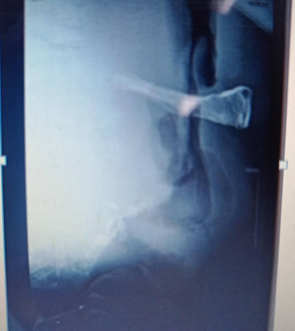
Huge retropharyngeal abscess almost abutting epiglottis
Plan:
Patient counselled and prepared for Incision and Drainage ± Tracheostomy.
In Operation Theatre
Found to be severely obstructed and resisted anaesthetist attempt to pre-oxygenate him or make him lie down for endotracheal intubation. He could not lie down for tracheostomy.
Next option : awake intubation- superior laryngeal nerve block was done bilaterally, followed by flexible nasolaryngoscopy, laryngeal inlet identified after several attempts, bougie inserted through vocal cords and rail-rolled endotracheal tube into the trachea. This stage took about 2 hours because patient was not cooperative and also technically challenging negotiating the swelling to get into the larynx.
After successful intubation, Boyle-Davies mouth gag was inserted and patient placed in tonsilliectomy position. Huge bulge on posterior oropharyngeal wall, more on the right, extending down to the hypopharynx. Cruciate incision was made .
Copious offensive mucopurulent fluids , about 500mls was drained. This was followed by direct laryngoscopy-larynx found to be normal but pus found in both pyriform sinuses. Following this, tracheostomy was done to protect airway and NG tube passed for feeding.
Postoperative Managements
- IV fluids-
- IV Augmentin 1.2 g 8 hourly
- IV Flagyl 500mg 8 hourly
- IV Paracetamol 1g 8 hourly
- NPO (Nil Per Oral)
- Started feeding per NG tube from POD1 .
- Patient progressed well. Ngtube removed on POD 7 and started feeding and tolerating per os.
- Successfully decanulated on POD 9.
- Discharged on oral medications on POD 10.
- Reviewed at OPD 2 weeks after discharge on POD 24 and was found to be doing well.
- Discharged from OPD to see for review PRN.
Conclusion
- The diagnosis of retropharyngeal can be challenging in both adults and children.
- A high index of suspicion is required.
- A simple soft tissue x-ray of the neck for any patient with dysphagia is an initial less expensive imaging modality that could help make the diagnosis.
- A competent interpretation of soft tissue x-ray of the neck is mandatory for emergency medicine physicians, family physicians, Specialty ENT doctors and ENT specialists.
Share Post On:
Recent Posts
-
Nuggets of ORL-OTOLOGY
-
Nuggets of ORL-RHINOLOGY
-
Nuggets of Otorhinolaryngology-Basic sciences
-
Anatomy of the Muscles of the Soft Palate
-
Ethmoidal Arteries Ligation for Epistaxis
-
Submucous Cleft Palate (SMCP)
-
Approach to Ligation of the External Carotid Artery
-
Approach to Managing a 3-Year-Old Boy with a Foreign Body in the nasal cavity.
-
Approach to Managing a 3-Year-Old Boy with a Foreign Body impacted in the ear canal.
-
Endoscopic Sphenopalatine Artery Ligation (ESPAL) for Epistaxis
-
Surgical Management of Epistaxis
-
Technique of Incision and Drainage of Septal Hematoma/Septal Abscess
-
Upper Aerodigestive Tract Foreign Body Impaction
-
Incision and Drainage of Hematoma Auris
-
Rigid Bronchoscopy for Retrieval of Foreign Bodies in Children
-
Foreign Body Impaction in the Larynx, Trachea, and Bronchi
-
Leadership Position is a Tool, not a Trophy
-
Carcinoma of the Oropharynx
-
Peritonsillar Abscess
-
Ethics of Doctor-Patient Relationship
-
Doctor-Patient Relationship Case Scenarios
-
Asymmetrical Tonsils and Approach to Evaluation and Management
-
Nasal Polyposis
-
Rigid Oesophagoscopy and Complication
-
Anatomy of Oesophagus
-
Stridor, Snoring, Stertor And Wheezing: How They Compare
-
Temporomandibular Joint (TMJ)
-
Otoacoustic Emissions
-
Tympanometry
-
Functional Endoscopic Sinus Surgery (FESS)
Categories
RELATED POSTS
Get in Touch
Read doctor-produced health and medical information written for you to make informed decisions about your health concerns.